 |
| About Bioline | All Journals | Testimonials | Membership | News |
|
||||||
|
||||||
Indian Journal of Human Genetics, Vol. 8, No. 2, Jul-Dec, 2002 pp. 66-68 Chromosomal Analysis in Recurrent Spontaneous Aborters Lakshmi Kalpana V, Satyanarayana M, Prabahker S and Sarvani R. Department of Human Genetics Andhra University, Visakhapatnam 530 003, Andhra Pradesh, India
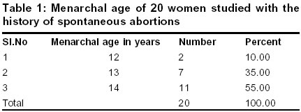
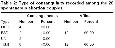
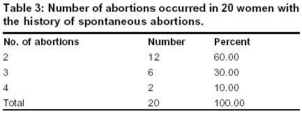
Code Number: hg02013 A type of endomitotic chromosome reduplication in the interphase stage of cell cycle was found in lymphocyte cultures of couples with spontaneous abortions. To find out the presence of this anomaly chromosomal analysis was performed in a series of 20 women with repeated spontaneous abortions and their husbands. Demographic data was also collected from them. Out of 40 individuals, a 27 years male whose wife has experienced three spontaneous abortions was found to have Endoreduplication. His wife was normal with 46, XX chromosomal complement. Key Words: Endoreduplication, Spontaneous abortions, Endomitotic chromosomes, Interphase chromosomes, Consanguinity, Gestation period, DNA replication, Aneuploidy. Introduction It is considered that about 15% of all recognized pregnancies terminate in spontaneous abortions1. The hypothesis advanced by streeter2, hertig & rock3 that the principal aetiological factor in spontaneous abortion is an intrinsic anomaly in the fertilized ovum, a so called germplasm defect and has been confirmed by the observation that a high proportion of such abortuses are chromosomally abnormal4. Earlier reports showed that 25 to 35% of all recognizable spontaneous abortions are caused by chromosomal anomalies57. The reported frequency of chromosomal abnormalities in embroys is 3.8 to 7.5%, in foetuses 1.5 to 2%, in still births 6.0 to 7.0%, in newborns 0.6%, in children (upto 7-8 years) 0.5% and in adults 0.4%.8 In adults, chromosomal abnormalities are found 4 in 1000, of which 50% occur in genetically balanced form. Of all spontaneous abortions 50 to 80% showed a chromosome anomaly & 94% of all detectable chromosomal abnormalities were associated with clinically recognizable fetal wastage. Chromosomal abnormality is a major cause of fetal loss and extensive surveys have been showed that 20-30 % abortuses are aneuploid.4,9,10 The mean incidence of cytogenetic anomalies in 6639 couples investigated for recurrent abortions was 6.65%. 11-13 Material and methods Twenty couples with recurrent spontaneous abortions were studied from visakhapatnam district. Majority of the cases had two or more spontaneous abortions. A complete case history was taken from the patient, including personal particulars, family history, detailed pedigree chart and the result of laboratory or other investigations. Chromosomal preparations were obtained using standard method and were subjected to gtg-banding. A minimum of 25 banded metaphase plates were analysed for routine karyotyping. Results Table 1 shows the menarchal age of the women studied with the history of spontaneous abortions. It is evident that 2 cases (10.00%) belongs to the 12years age group. 7 cases (35.00%) fall in the 13 years age group and 11 cases (55.00%) were into the 14 years age group when the age at attaining menarche was considered. The menarchal age is high in more number of women with the history of spontaneous abortions. In the present study, 12 cases (60.00%) were of affinal marriages; remaining 8 cases (40.00%) were consanguineous. Out of these 8 cases, 4 (20.00%) fall under mother's brother's daughter, 2 (10.00%) under father's sister's daughter and the remaining 2 (10.00%) under unknown categories of consanguinity (table 2). The higher rate of consanguinity in the present study could be due to small sample size. Table 3 shows, 12 cases with 2 (60.00%) abortions, 6 cases with 3 (30.00%) abortions, and 2 cases with 4 (10.00%) abortions. It has been observed that more number of couples are having two spontaneous abortions. Discussion The demographic approach is essential for the proper measurement of selective differences in human populations. Demographic processes are direct manifestations of physiological and biological parameters. Factors like menarche, menopause, gestation period, consanguinity regulate the demographic structure of a population to a considerable extent. The age of menarche varies to some extent with genetic background like family history, geographic location socioeconomic status, nutrition build life style. According to Tanner's studies,13 the menarchal age varies between 10-16 years with a average of 13.5 years. Abortion or the reproductive wastage is defined as the expulsion or extraction from its mother of an embryo. It is generally accepted that 15% of all recognised pregnancies terminate in spontaneous abortions. The importance of human reproductive wastage has been increasingly emphasized during recent years. Various studies have confirmed that the total frequency of chromosomal anomalies is 50 to 60%. The highest frequency of chromosomal defects is seen in early spontaneous abortions. Earlier it has been reported that the frequency of chromosomal abnormalities ranged from 0.86% - 45%. The upper limit was reported by Nordenson14 and Gupta et al15 and the lower limit was reported by Kajii et al.16 The high variation in percentage of abnormal cases in these surveys may be attributed to the lack of uniformity in selecting the couples i.e., those with different number of spontaneous abortions (one or more spontaneous abortions ) and with different gestational ages. In the present study out of 40 cases, a 27 years male, with 3 repeated spontaneous abortions of his wife showed endoreduplication. Out of 3 pregnancies, 2 of which ended at about 2 months of gestation and 1 at about 3 months of gestation. The proband was born when her father and mother were 32 years and 24 years respectively and no abnormalities were noticed at birth or thereafter. He was normal phenotypically and mentally, with a height of 168 cms and a weight of 75 kgs. The wife of the proband aged 25 is a healthy and well developed female with normal 46, XX chromosomal complement. Metaphase analysis of the male showed 6% cells with endoreduplication. Endoreduplication refers to the process by which chromosomes go through two or more dna replication cycles but fails to undergo cell division. A type of endomitotic chromosome reduplication in the interphase stage of cell cycle. Endoreduplication takes place without any mitosis like manifestation and consists of a double, triple or multiple reduplications of interphase chromosomes. This leads to the mitotic disturbances finally resulting in aneuploidy. In 1990, Murthy & Prabhakara17 reported a female with history of spontaneous abortion and subsequent birth of Down syndrome child. Her chromosomal analysis revealed 46, XX with pericentric inversion of 9qh, while her husband's was normal. Metaphases analysis of the female showed 20.5% cells with premature centromere division, 4% cells with endoreduplication and 2% with polypolidy. These frequencies were considerably higher as compared to a normal control. They concluded that inv (9qh) might have some inter chromosomal effect leading to higher incidence of mitiotic disturbances finally resulting in aneuploidy. This predisposition is evident by spontaneous abortion and Down syndrome child. Hence, the observation in the present study showed that patients with endomitotic chromosome reduplication in the interphase stage of cell cycle may predispose the cell division errors due to chromosome instability and thus may lead to spontaneous abortions. Such a correlation awaits a better understanding of the nature of these cytogenetic observations and the events controlling mitosis and meiosis at the molecular level. References
Copyright 2002 - the Indian Society of Human Genetics The following images related to this document are available:Photo images[hg02013t3.jpg] [hg02013t2.jpg] [hg02013t1.jpg] |
| |||||||||
