 |
| About Bioline | All Journals | Testimonials | Membership | News |
|
||||||
|
||||||
Indian Journal of Human Genetics, Vol. 9, No. 1, Jan-Jun, 2003, pp. 21-24 Radiation-induced chromosomal hot spots at G1 and G2 stages of human lymphocytes in culture R. Murugesan Department of Biotechnology, Kasturba Medical Colleage, MAHE,
Manipal-576119, India.
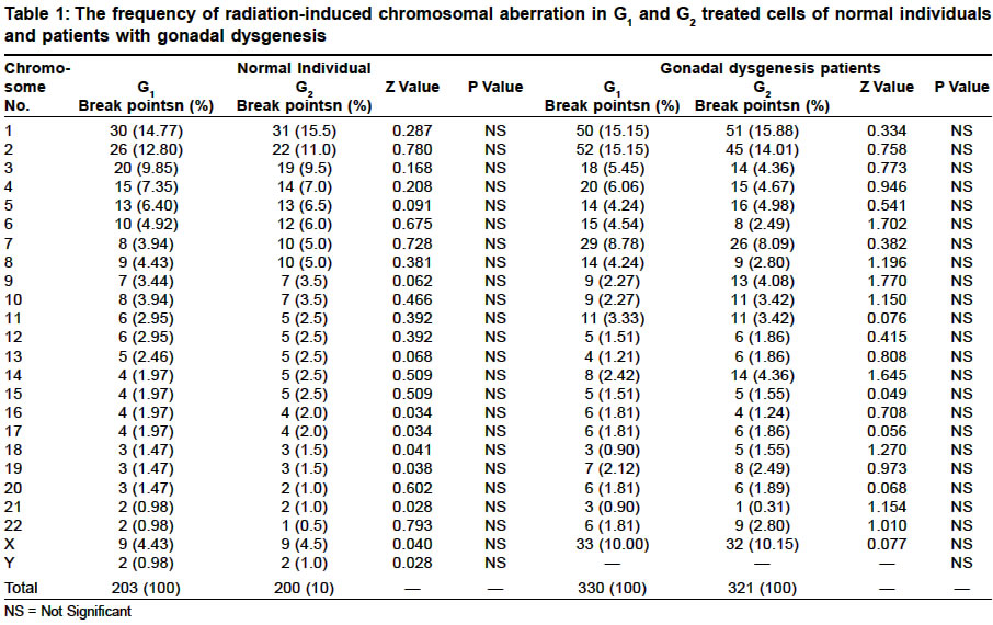
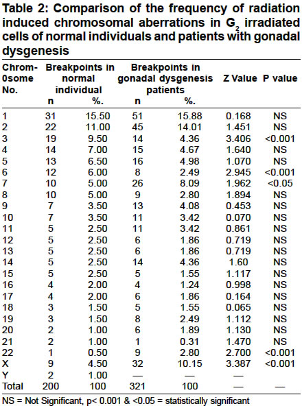
Code Number: hg03006 Radiation-induced chromosomal break points in cultured lymphocytes of normal healthy individuals as well as of those with certain genetic disorders are reported to be localized at certain specific loci (hot spots). These reports are based on studies carried out in lymphocytes irradiated at G1 stage. The present study examines whether the location of hot spots and the frequency seen in cells irradiated at G1 are similar to those irradiated at G2 stage of the cell cycle and also tests whether cells of patients exhibit hot spots on irradiation.The results showed that the radiation induced chromosomal break points to be similar in those irradiated are G1 and G2 stages of the cell cycle and also that cells of patients exhibited chromosomal hot spots. Key Words: Radiation, Break points, Hot spots. INTRODUCTION The chromosomal break points, more frequently observed in certain region of chromosomes are called as mutational "hot spots". Knehr et al6 reported that radiation- induced chromosomal rearrangements are non-random and they cluster at specific sites within specific chromosomes. Kano and Little2 had earlier observed non-random distribution of translocation break points in X-irradiated human diploid fibroblasts. Borrios et al3 examined the distribution of break points in lymphocytes from cancer patients and band 1q32 was proposed by them as a hot spot. Sugunasankari4 applied low dose of radiation and identified specific break points on chromosome 7q22 and Xq22 in patients with gonadal dysgenesis. Employing a similar procedure, Murugesan5 located specific chromosomal break points in cells of individuals with haemophilia, ichthyosis, and retinitis pigmentosa, and observed specific chromosomal hot spots for these genetic disorders. The break points are thus considered as specific to certain diseases. Interphase can be partitioned into three phases namely G1, S and G2 in mitotically proliferating cells. Blood samples of the patients as well as normal individuals are irradiated mostly at G1 and the chromosomal aberrations get repaired when they pass through S phase. However, in blood samples irradiated at the very late G2 or at early prophase and fixed 45 minutes after irradiation, all the chromatid aberrations will remain as such and will be available for analysis. This study was carried to find out the intra-and inter-chromosomal radiosensitivity in human chromosomes treated at G1 and G2 stages and to study the genetic control of radiation-induced chromosomal aberrations with special reference to the chromosomal hot spots in healthy individuals and in patients with gonadal dysgenesis. MATERIAL AND METHODS Chromosomal preparations from the cultured lymphocytes of five healthy individuals and five patients with gonadal dysgenesis were obtained according to the modified method of Hungerford.6 The radiosensitivity of the lymphocytes of patients and control set of individuals was assessed by exposing the cultured lymphocytes to gamma radiation 60Co, at Radiography laboratory, MERADO, CSIR Complex, Chennai. The lymphocytes were irradiated at a dose of 2Gy at G1 phase (dose rate: 14 cGy/minute) before adding mitogen (PHA). The cultures were incubated at 370C for 72 hours. For the irradiation at G2 phase, cultured lymphocytes at 691/2 hours, were irradiated at a dose of 0.7Gy (dose rate: 14cGy /min), and colchicine was added immediately after irradiation to these cultures and incubated at 370C for 25 minutes. The slides prepared were stained as per the procedure of Marimuthu et al.7 Fifty well spread metaphases were analysed to record various structural abnormalities from G1 and G2 treated cells and the frequencies and the location of the break points were recorded for each chromosome. RESULTS The frequency of chromosomal aberrations induced by radiation in each of the twenty-four chromosomes was found to be similar in cells irradiated either at G1 or G2. A similar frequency was recorded also in the cells of genetically defective individuals (Table 1). However, the incidence of aberrations in specific chromosomes of normal and genetically defective individuals differed and the frequency was found to be higher in the latter group (Table 2). From the data (Table 2) it is evident that the response of chromosomes 7, 22 and X of patients was significantly different compared to those of healthy individuals. DISCUSSION Reports are available on radiation-induced non-random chromosomal break points in normal individuals as well as in patients with genetic disorders.1,2 Non-random distributions of translocation breakpoints indicate the existence of hot spots or sites. Specific chromosomes exhibiting frequent involvement in rearrangements were reported by Kano and Little2 in X-irradiated human diploid fibroblasts. Increased instances of translocation in the short arm of chromosome 1 (1p22) are reported and this is the most commonly involved chromosome in human cancer.8 Patients with gonadal dysgenesis exhibited significant difference in break points on chromosomes 3, 6, 7, 22 and X. While chromosomes 3 and 6 showed decrease, increase was observed in chromosomes 7, 22, X. The involvement of X-chromosome in gonadal dysgenesis is on record.9 Natarajan10 reported that radiosensitivity differs when cells of ataxia telangiectasia patients irradiated at G1 and G2. Hsu et al11 observed frequencies of chromatid aberrations amounting to at least three times higher in Chinese hamster fibroblasts irradiated during G2 compared to those irradiated at G1. In the present study frequency of chromosomal break points are similar both in G1 and G2 stages even when the radiation dose given at G2 is less than one third of that at G1 stage. The pattern of distribution of chromosomal aberrations on various chromosomes in the genome is similar in cells irradiated at G1 and G2. Irradiation of cells at G2 provides more reliable picture for a comparative study of the radiosensitivity or hot spot analysis of individual chromosomes between control individuals and in patients with genetic disorders. CONCLUSIONS The radiosensitivity of individual chromosomes and the location of the radiation-induced chromosomal break points are found to be genetically controlled. There exists an intra and inter-chromosomal variation in the production of radiation-induced chromosomal aberrations. Hot spots are observed on specific loci of a given chromosome. The frequency and location of the radiation-induced breakpoints are similar both in the G1 and G2 irradiated leucocytes. However, radiosensitivity of lymphocytes of patients with gonadal dysgenesis is significantly higher than that of the normal individuals in G1 and G2 treated cells. A broad correlation was observed between the length of the chromosome and the frequency of chromosomal aberrations in cells of normal individuals, though this was not observed in cells of patients with gonadal dysgenesis. ACKNOWLEDGEMENTS The author expresses his gratitude to Prof. K. M. Marimuthu and Prof. P. M.Gopinath for advice and guidance. He is thankful to CSIR for financial support. The work was carried out at the Department of Genetics, Dr A.L.M.P.G.I.B.M.S, University of Madras, Taramani, and the author thanks the University of Madras for facilities. REFERENCES
Copyright 2003 - the Indian Society of Human Genetics The following images related to this document are available:Photo images[hg03006t2.jpg] [hg03006t1.jpg] |
| |||||||||
