 |
| About Bioline | All Journals | Testimonials | Membership | News |
|
||||||
|
||||||
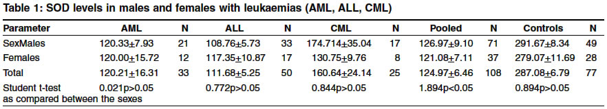
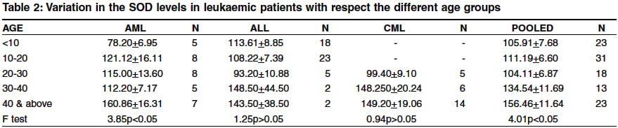
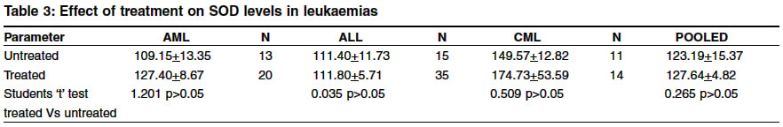
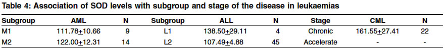
Indian Journal of Human Genetics, Vol. 10, No. 1, Jan-June, 2004, pp. 9-12 Original Article Quantitative variation of superoxide dismutase levels in leukaemias Poongothai AR, Vishnupriya S, Ragunadharao D Department of Genetics, Osmania University, Hyderabad Code Number: hg04003 ABSTRACT Superoxde dismutase is dimeric antioxidant enzyme responsible for quenching of superoxide radicals which are released during the chemical reactions of the various metabolic pathways .The enzyme levels of SOD are altered to a considerable extent in various diseased states exhibiting either elevation or depletion in their enzymatic activity.As this phenomenon was found to be more evident in leukaemias, we have estimated the Cu-Zn SOD levels in the red cells of the leukaemic patients in order to evaluate the efficiency of anti oxidant system and its relation to leukaemiogenesis. The overall mean SOD levels in the leukaemic patients (AML, ALL and CML) were significantly low (124.97±6.46) as compared to that of the normal controls (287.08±6.79). In respect to the age, the mean SOD levels were found to be elevated indicating the extent of the free radicals might have stimulated the production of the antioxidants.In response to chemotherapy, the mean SOD levels were observed to be elevated but not the extent of the normal levels among the treated groups in AML(127.40±8.67) and CML (174.73±53.59) types of leukaemias as compared to that of the untreated groups. In general the stage of leukaemia did not cause any variation in the SOD levels. However, the L2 in ALL (107.49±4.88), blast crisis in CML (154.00±19.86) showed reduced levels of SOD.INTRODUCTION Superoxide dismutase is dimeric antioxidant enzyme responsible for the quenching of superoxide radicals which are released during the chemical reactions of the various metabolic pathways. There are several ways by which free radicals are formed.Inside the body, oxygen as well as hydroxyl radicals are generated during energy productions.Once formed, these free radicals initiate their own reactions thereby exerting potentially harmful effects on the various systems of the body.In the normal course, the free radicals are converted to a less reactive form (H2O2) by the free radical quenching enzymes (SOD) which are naturally found in the mitochondria. This enzyme catalyzes the removal of the O2 free radical, thus protecting the O2 metabolizing cells against the harmful effects of the superoxide free radicals. However, the enzymatic levels of SOD are altered to a considerable extent in various diseased states exhibiting either elevation or depletion in activity.[1] As this phenomenon was found to be more evident in leukaemias, Cu-Zn SOD levels were estimated in the red cells of the leukaemic patients in order to evaluate the efficiency of anti oxidant system and its relation to Leukaemiogenesis. Similar studies were carried out by Devi et al[2] in 30 different untreated leukaemic patients. MATERIALS AND METHODS Blood samples were collected from 108 patients, with confirmed diagnosis of leukaemia, reporting to Nizams Institute of Medical Sciences (NIMS), medical oncology section Hyderabad, India. The patient population is representative of the general population in terms of socioeconomic condition and ethinic diversity. However, the variation in SOD levels of leukaemic patients with respect to their sex, age, stage and treatment of disease are discussed in this paper. The patients were divided into groups based on their sex, the type of leukaemia (ALL, CML, AML)and mode of treatment (treated, untreated). The clinical findings (diagnosis, complete blood picture, stage of the disease and therapy to be/being followed) were recorded with the help of medical oncologist. In addition, blood samples were also obtained from 77 healthy age and sex matched randomly selected individuals from general population to serve as controls. Detailed information regarding previous medical history, nutritional status and parental consanguinity was obtained from patients and controls through personal interview using a prescribed proforma. Superoxide dismutase levels were estimated in the red cells of the leukaemic patients as described earlier.[3] SOD levels were expressed as mg/liter. The significance between different groups was tested using student t - test or F test.The level of significance was at p<0.05. RESULTS AND DISCUSSION The overall mean SOD levels of leukaemic patients (AML,ALL and CML) were significantly low compared to that of the normal controls, the reduction being more prominent in ALL [Table - 1]. Similar findings of depletion in antioxidant - superoxide dismutase levels have also been reported earlier in other cancers[4],[5],[6],[7],[8],[9] indicating the role of antioxidants in the prevention of cancer. Senturker et al[10] studied the antioxidant enzyme levels and oxidative damage in lymphocytes of children with acute lymphoblastic leukaemia (ALL) and also in disease free children (patients) and they observed that a higher levels of DNA base lesions and reduced antioxidant levels in former group as compared to the latter. This might indicate a possible link between decreased activities of antioxidant enzymes and increased levels of base lesions due to damaging effect of free radical reactions released during oxidative stress leading to the carcinogenic process. A increased concentrations of thiobarbituric acid reacting substance (TBARS) were found in spite of antioxidant defense activation suggesting that cellular peroxidative reactions might be occurring during the carcinogenic process.[11] From the above findings, it could be concluded that elevation or depletion in the SOD levels could be due to the cause of or the consequence of the disease onset and progression. The significant sex difference in SOD levels were observed only in CML patients, where males showed higher level of enzyme activity as compared to females [Table - 1]. Earlier, it was observed that total antioxidant status (TAS) was lower in females than in males, however, there was significant decrease in TAS levels with age in male but not in female indicating the genetic difference found in management of oxidative status.[12] This might explain the significant sex difference found in SOD enzyme activity in present study in the case of CML. Both types of acute leukaemias (AML and ALL) have exhibited reduced SOD levels, though sex difference was not apparent, as compared to controls indicating failure of antioxidant mechanism to counteract the extensive oxidative damage resulting in leukaemia which progresses aggressively in short time. In the case of CML, the SOD levels are higher than that of the acute leukaemias which supports our previous contention of defective antioxidant mechanism and fast progression of disease in the case of acute leukaemias. [Table - 2] shows the variation of SOD levels in the leukaemic patients with different age groups. In the age group between 0-10 years and 10-20 years there is a sudden increase in the mean SOD levels of AML patients (78.20±6.95 to 121.13± 16.11). Whereas in the age group between 20-30 years and 30-40 years, there is a decline in the mean SOD levels. There was no significant trend in SOD levels with respect to age in both ALL and CML. Various studies have reported the process of aging associated with the degree of the antioxidant activity. Casado et al[13] reported the mean SOD levels were increased in the diseases of the aged individuals such as cardiovascular diseases ,myomas, chronic obstructive pulmonary disease and acute cerebral accident whereas in osteoporosis diseases, activity of SOD remained standard, but the SOD enzyme levels were seen to be decreasing with the process of aging. Whereas in another report[14] it has been shown that the excessive production of free radicals in the organism and the imbalance between the concentrations of these and the antioxidant defenses was related to the processes such as aging and the development of several diseases such as cancer. The effect of treatment on SOD levels in leukaemic patients has been shown in [Table - 3]. The mean SOD levels were observed to be elevated but not to the normal level among the treated patients as compared to that of the corresponding untreated patients of the AML and CML, indicating the response to chemotherapy. The subgroup of leukaemia did not cause any significant variation in the SOD levels in the AML. However L2 subgroup in ALL and blast phase in CML have shown reduced level of SOD enzyme [Table - 4], indicating inefficient antioxidant mechanism to combat the severity of the disease. Similar observations were reported by Zhu et al[15] where the SOD activity declined in the blast phase of the acute leukaemia but increased considerably to normal levels during remission suggesting the SOD enzyme might be playing a protective role during the onset and progression of leukaemia and can serve as useful parameter in diagnosis and for clinical course of the Leukaemiogenesis. ACKNOWLEDGEMENT We are thankful to the medical and the non technical staffs of Nizams institute of medical Sciences in helping us through the data collection The financial assistance provided by council of Scientific and industrial Research (CSIR), Rameswardas birla Smarak Kosh and Indian Medical association (Mediciti Hospitals, Hyderabad) is greatly acknowledged. REFERENCES
Copyright 2004 - Indian Journal of Human Genetics The following images related to this document are available:Photo images[hg04003t3.jpg] [hg04003t1.jpg] [hg04003t4.jpg] [hg04003t2.jpg] |
| |||||||||
