 |
| About Bioline | All Journals | Testimonials | Membership | News |
|
||||||
|
||||||
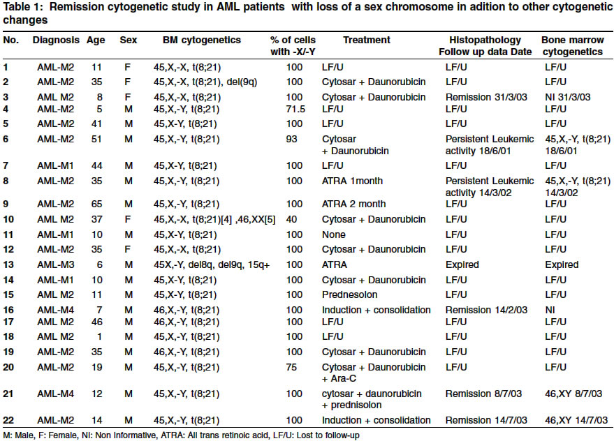
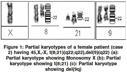
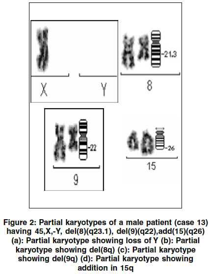
Indian Journal of Human Genetics, Vol. 10, No. 1, Jan-June, 2004, pp. 22-25 Original Article Loss of sex chromosome in acute myeloid leukemia Bakshi Sonal R, Kakadia Purvi M, Brahmbhatt Manisha M, Trivedi Pina J, Rawal Shwetal M, Bhatt Samarth S, Parikh Bharat J, Patel Kirti M, Shukla Shilin N , Shah Pankaj M Cell Biology Division, Division of Research, The Gujarat Cancer Society, Department of Cancer Biology, The Gujarat Cancer and Research Institute, NCH Campus, Asarwa, Ahmedabad - 380 016 Code Number: hg04006 ABSTRACT Loss of sex chromosomes has been reported in normal and malignant marrows and its frequency increases with age in both situations. It is not clear whether the sex chromosome loss is a critical mutational event for neoplastic transformation or a genetic change related to ageing. The present study was undertaken to analyze incidence of loss of sex chromosomes in leukemia patients. Karyotypic analysis in bone marrow cells was carried out in total 270 AML patients registered at G.C.& R.I. during January 2000 to October 2003. Out of 270, 22 patients had loss of sex chromosome in addition to other disease specific chromosomal abnormalities. Out of 22 patients, 50% (11 of 22) were of the pediatric age (up to 14 years), and only 10% (3 of 22) patients were above the age of 50 years, maximum age being 65 years. On follow-up, only in patients with pathological remission normal 46XX/XY karyotypes were seen. Whereas in patients with persistent leukemic activity, clones with loss of sex chromosome were observed. The results indicate that sex chromosome loss in these cases may be equivalent of a clonal cytogenetic process rather than related to ageing process.INTRODUCTION In normal aging males, the loss of Y chromosome in bone marrow cells has been appreciated since the early 1970s; subsequently the loss of this chromosome was reported to occur in a variety of hematological disorders.[1],[2],[3],[4],[5] In females, a corollary loss of X chromosome also occurs with advancing age.[6] The frequency of loss of Y chromosome in males with no evidence of hematological disease has been reported to be 7.7%.[7] Cells with - Y are observed more often in males over age 55 than in younger males. The frequency of - Y cells increases with advancing age and is significantly greater in case with MDS, MPD, ANLL, or LPD than in subjects with no evidence of disease, the later rarely exhibit more than 75% of cells with 45, X, -Y. Specific chromosomal abnormalities are well recognized in acute leukemia, and are emerging as major determinants for treatment, response and survival.[8],[9] Loss of Y chromosome as a sole abnormality or with additional cytogenetic changes has been reported in acute myelogenous leukemia (AML), although it is not clear whether or not this abnormality is a marker for the leukemic clone.[10],[11],[12] The significance of sex chromosome loss as the only karyotype anomaly in hematological disorders has not been clearly established.[11] Studies have supported the theory that Y loss is a nonphenotypic event associated with the aging process in males.[13] It has been reported that in 2 AML patients the lost X and Y (individually) chromosomes reappeared after therapy and during clinical remission. Here we present a retrospective study of remission cytogenetics in AML patients with loss of a sex chromosome in association with other chromosomal abnormalities. MATERIALS AND METHODS Between January 2000 and October 2003, pretreatment bone marrow specimens of 270 patients with AML registered at The Gujarat Cancer and Research Institute (Ahmedabad) were received for cytogenetic analysis. There were 178 males and 92 females and their age range was 1-75 years. The bone marrow samples were collected in Na-Heparinized vaccuetainer containing RPMI-1640 medium with antibiotics and standard short-term cultures were carried out in RPMI-1640 medium supplemented with serum, heparin and antibiotics at 37°C in 5% CO2 incubator. Harvesting and GTG banding were carried out according to standard procedures, followed by karyotyping as per the ISCN 1995 guidelines.[14] The automatic karyotyping system by Zeiss and IKAROS software from Metasystems, Germany was used. The patients were followed-up for karyotypic analysis during the course of treatment. RESULTS The pretreatment bone marrow cytogenetic study revealed that out of 270 patients included in the study, 22 patients had loss of a sex chromosome in addition to other disease specific chromosomal abnormalities [Table - 1]. Out of 22, 20 had t(8;21) , one had t(8;21),del(9q) [Figure - 1] and one had complex karyotype with del(8q),del(9q),add15q [Figure - 2]. Out of these 22 patients, 50% (2 females and 9 males) were under 14 years, 40% (3 females and 6 males) were between the age of 14 & 50 years, and 10% (2 males) were above 50 years. Of the 22 patients, fifteen patients were lost to follow up and the one with complex karyotype expired during the course of treatment. Among the remaining 6; post treatment bone marrow examination showed that in the cases where morphologically persistent leukemic activity was seen, cytogenetics showed persistent loss of sex chromosome in addition to t(8;21); and in cases where marrow showed morphological remission, cytogenetics revealed reappearance of normal 46,XY clone [Table - 1]. DISCUSSION In the present report out of the 22 patients 21 patients showed loss of a sex chromosome as a secondary anomaly to t(8;21) and one showed complex karyotype with -Y who expired during the treatment. It has been reported that 18% of all AML with an abnormal karyotype show t(8;21); moreover, -Y, -X and del(9q) appear as a secondary anomaly at a frequency of 39.3%, 16.3% and 12.7% respectively.[15] The two cases where complete pathological remission was seen, bone marrow cytogenetics showed normal diploid karyotypes and in the two cases where pathologically persistent leukemic activity was seen, there was persistent loss of sex chromosome in proliferating clones. The present results indicate that the sex chromosome loss may be equivalent of a neoplastic clone rather than related to aging process, as patients included in the present study were of the lower age group as compared to the literature.[16] In the present study t(8;21) serves as an internal control as loss of sex chromosome as a sole anomaly in a few metaphases could have been easily attributed to the aging phenomenon rather than indicative of the disease. Possible significance of loss of Y chromosome in neoplasia have been postulated as; Y chromosome harbors a tumor suppresser gene, which when lost or modified, gene(s) presumably located on the X chromosome may be affected leading to abnormal proliferation.[16], [17] Further studies are required for better understanding of the role of sex chromosome loss in hematological neoplasia. Frequency of loss of sex chromosome should be estimated from normal tissue also in addition to leukemic cells. Comparison of age, clinical course, and percentage of cells with -X or -Y by interphase fluorescence in situ hybridization may give better comparison of the baseline frequency of loss of sex chromosome among normal subjects and leukemia patients. ACKNOWLEDGEMENTS The authors would like to acknowledge the valuable technical support of Ms. Sheeba John.REFERENCES
Copyright 2004 - Indian Journal of Human Genetics The following images related to this document are available:Photo images[hg04006f2.jpg] [hg04006f1.jpg] [hg04006t1.jpg] |
| |||||||||
