 |
| About Bioline | All Journals | Testimonials | Membership | News |
|
||||||
|
||||||
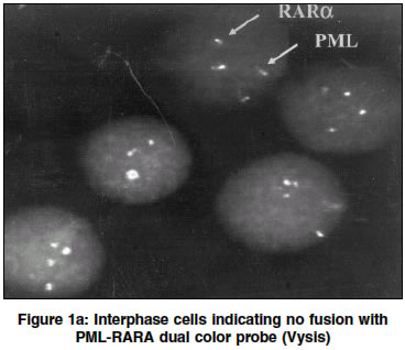
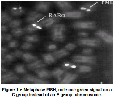
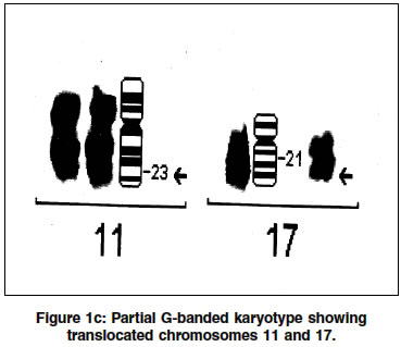
Indian Journal of Human Genetics, Vol. 10, No. 1, Jan-June, 2004, pp. 26-28 Letter To Editor Fluorescence in situ hybridization studies: Break apart or not? Bakshi Sonal R, Brahmbhatt Manisha M, Trivedi Pina J, Rawal Shwetal M, Kakadia Purvi M, Bhatt Samarth S Cell Biology Division, Department of Cancer Biology, The Gujarat Cancer & Research Institute, NCH Campus, Asarwa, Ahmedabad - 380016 Code Number: hg04007 Sir, We would like to share our experience with acute promyelocytic leukemia (APML) molecular cytogenetic studies. Recent debate regarding interpretation of FISH (Fluorescence in situ hybridization) with BCR/ABL (ES) probe for detection of der(9q) deletion in t (9;22)(q34;q11.2) published in the journal Cancer Genetics and Cytogenetics[1],[2] was interesting for the cancer cytogenetics community using commercially available FISH probes. Acute Promyelocytic leukemia (M3 in the FAB classification) is specifically associated with the t(15;17)(q22;q21) resulting in PML/RARá fusion gene, a chromosomal variant that shows a good response to all-trans retinoic acid (ATRA) therapy.[3] Variant APML cases involving RARá but not the PML gene have been reported. Cases with translocation t(11;17)(q23;q21), which fuses the PLZF and RARa genes do not respond to ATRA therapy. In contrast, cases with t(11;17)(q13;q21)and t(5;17)(q35;q21) which bring about fusion of RARA with NuMA and NPM, respectively, are reported to be sensitive to ATRA.[3],[4] A study of identification of APML patients with variant translocation has been initiated at The Gujarat Cancer & Research Institute since December 2000. Conventional cytogenetics and FISH analysis using dual color probe for PML and RARá genes (32-191009, Vysis) are carried out at Cell Biology Division. Presence of two green and 2 orange spots depict no PML-RARá gene fusion, whereas 1 green, 1 orange, and 1 yellow spots signal specific fusion. In one sample of cultured bone marrow cells, FISH analysis of interphase and metaphase revealed no PML-RARá gene fusion [Figure - 1]a,b,c. Interpreting absence of PML-RARá gene fusion needs close examination in such cases (see below). The RARa signal was observed on a C group, instead of an E group chromosome suggesting variant translocation. Possible involvement of chromosome 11 was confirmed using whole chromosome paint FISH for chromosome 11 (Vysis) as FISH probe for 11q23 was not available with us. Cytogenetic results revealed later, t(11;17)(q23;q21) [Figure - 1]d, an APML subtype that is resistant to ATRA therapy. In light of the variants reported, a new RARá DNA probe tagged with adjacent green & orange fluorescent dyes has been launched recently (32-191011, Vysis), obviously, not to miss any of the variant RARa rearrangements. Two yellow/fusion signals indicate no RARa gene rearrangement; whereas, one fusion signal and one pair of split signals (an orange and a green signal) indicate RARa gene translocation. Now, if sample has no analyzable metaphases, which is likely due to several reasons, or when FISH and simultaneous banding in metaphase is not possible, the translocation partner of RARá is not known. The new break-apart probe though useful, will not identify partner chromosome site involved in RARá rearrangement always, thus identification of ATRA-responsive or non-responsive variant may still remain a question. Looking at the price of the commercially available FISH probes, it is worth considering these aspects before ordering each and every probe available for every possible DNA strand reported to be involved in a malignancy. Theoretically, in case of a leukemia patient candidate for bone marrow transplantation, a very sensitive pre-therapy evaluation may require 2 probes each for 7q and 5q, and 4 probes for the regions encompassing 13q! Similarly, for RT-PCR one has to carefully choose the right primers. To summarize the situation of a cancer cytogeneticist, conventional cytogenetic analysis is a must in identifying hitherto unknown chromosomal abnormalities (that, if recurrent, leads to a new FISH probe marketed!), on the other hand, its limitations with submicroscopic genetic rearrangements, and frequent dearth of analyzable metaphases necessitates molecular cytogenetics. Molecular cytogenetics in turn, offers greater sensitivity with known marker(s), but is never enough to get the whole picture! Gene microarrays may be the final answer, but until then we need to FISH carefully and give reports with appropriate disclaimers. ACKNOWLEDGEMENTS The authors are thankful to Dr. Pankaj M. Shah, Director, G. C. & R.I. for providing necessary facilities. Mr. Shailesh Patel, and Ms. Sheeba John are acknowledged for their valuable technical help.REFERENCES
Copyright 2004 - Indian Journal of Human Genetics The following images related to this document are available:Photo images[hg04007f1a.jpg] [hg04007f1b.jpg] [hg04007f1d.jpg] [hg04007f1c.jpg] |
| |||||||||

![Figure - 1]d](/showimage?hg/photo/hg04007f1d.jpg)