 |
| About Bioline | All Journals | Testimonials | Membership | News |
|
||||||
|
||||||
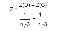
Indian Journal of Human Genetics, Vol. 11, No. 3, September-December, 2005, pp. 149-153 Original Communication Fluctuating asymmetry in dermatoglyphics of non-insulin-dependent diabetes mellitus in Bangalore-based population Ravindranath R, Joseph AM, Bosco SI, Rajangam1 S, Balasubramanyam V Department of Anatomy,St John's Medical College, Bangalore-560034, India Code Number: hg05029 Abstract BACKGROUND: The aetiology of NIDDM is believed to be as a consequence of genetic and environmental factors that impair metabolism. While little can be done on the genetic component, much can be done as a preventive measure in NIDDM. Because nothing much can be done prenatally, researchers have resorted to studying physical variables like dermatoglyphics (DGs). Dermatoglyphic patterns form on the finger pad and the palm prenatally and remain unchanged throughout life, thus these features may serve as markers for fetal origin of adult disease like NIDDM. Thus the concept of fluctuating asymmetry (FA) which has been defined as random differences between the right (R) and left (L) sides of a morphological trait has gained prominence in diseases like schizophrenia. When the distribution of R-L differences in a population sample approximates a normal curve with a mean approximately equal to zero, the variance of distributions of R-L difference is a measure of FA. Studies have shown that genetic factors may also have a link to FA in finger and a-b ridge counts. No studies have been reported on FA in NIDDM. FA derived from quantitative parameters in DGs of NIDDM may throw light on fetal origins of an adult disease. Hence this study has been undertaken.AIM: The present study aims at deriving FA from quantitative parameters in DGs of NIDDM compared to controls in the Bangalore based population. MATERIALS AND METHODS: Bilateral rolled finger and palm prints of 150 NIDDM patients (Males - 75, Females - 75) were compared to 120 controls (Males - 60, females -60) from Bangalore based population. FA measures derived from quantitative parameters (finger ridge counts, a-b ridge counts, main line index and palmar angles) were analysed. RESULTS: Comparisons were made in all parameters between homologous fingers of both hands using Pearson's product moment correlation coefficients (r). The difference in correlation coefficients between cases and controls was calculated using Fisher's Z transformation. 1-r2 an estimate of error variance thus measures FA. FA measures were significantly higher in NIDDM males for the 5th finger (FA=2.04) and for the palmar angle 'dat' (FA=2.24); for the NIDDM female a high FA was found in the 2nd finger (FA=2.17) compared to controls. CONCLUSION: Overall measures of the above ridge counts and angles and their derived measures of FA were prominent features of NIDDM in this sample. Keywords: Dermatoglyphics, fluctuating asymmetry, type 2 diabetes mellitus Circumstances during gestation may affect subsequent adult metabolism. A baby of reduced birth size, for example, carries an increased risk of insulin resistance in later life.[1] The mechanisms that underlie this prenatal, metabolic programming have not been established, and there is uncertainty about precisely when in gestation a fetal insult must occur in order to affect adult health.[2] Some reports suggest a relatively early time window in which pertinent gestational exposures may occur.[3] If an adverse fetal exposure were to occur before the twentieth week, we hypothesized that evidence of this effect must be found in the fingerprints of the offspring. A human fingerprint is the representation of dermal ridges on each fingertip. These dermal ridges are formed during gestational weeks 17 to 19.[4] On each fingertip, the ridge count provides a measure of fingertip growth activity during the early fetal period.[5] Several authors have studied finger tip patterns[6],[7],[8],[9],[10] and ridge count.[7] They[7] have found an increase in total finger ridge count in male diabetics and an increase in the t-d ridge count in both sexes. Main line index (MLI) has been studied on an unsegregated combination of juvenile and MODM.[8] Earlier studies have also reported dR45 (difference in ridge counts between the right fourth and fifth fingers) as a marker of early fetal metabolic programming.[11] However fluctuating asymmetry (FA) has not been reported for NIDDM in literature. FA is defined as random differences between the right and left sides of a morphological trait. When the distribution of the right minus left (R-L) differences in a population sample approximates a normal curve with a mean zero or close to zero, the variance in distributions of R-L difference is a measure of FA. While FA requires that R-L differences are random and non-directional, directional asymmetry (DA) involves a significant departure from zero to the normally distributed mean of R-L differences. Examples in humans include the asymmetry of bronchi and some internal organs and structures. There is evidence demonstrating rightward DA in finger ridge counts.[12],[13] Some authors emphasize that genetic factors have a weak link to FA in finger ridge counts[13] and a-b ridge counts.[14] It has been proposed that the degree of FA of an organism reflects the ′developmental instability′ of that organism.[15] The authors hypothesize that this developmental instability may be reflected in a marker if any as FA. FA can be ascertained at low cost, and therefore provide a convenient marker for studying the early gestational influence on chronic adult disease. The aims of the present study were: 1.To find out the quantitative parameters such as ridge count of individual fingers of right and left hands, sub total finger ridge count of each hand, a-b ridge count, main line index (MLI) and palmar angles(′ atd′, ′dat′ and ′adt′ angles) of each hand in controls and NIDDM patients. Material and Methods A total of 150 NIDDM patients (75 M and 75 F) from a catchment centre consisting of diabetic clinic from Specialists center and from St John′s Medical College, Bangalore along with 120 controls (60 M and 60 F) were studied. Their ages were 38 to 82 years and approximately 85 percent of subjects were outpatients. A screening questionnaire excluded participants with diseases or congenital abnormalities believed to be associated with fingerprint or other dermatoglyphic abnormalities. Patients were asked to wash their hands with soap water, so as to remove any oil or dirt. After written consent was given, finger prints were obtained by the ink and paper method[16] and finger and palm prints counted. Ridge counts for each finger tip were calculated from the number of primary dermal ridges that intersected or touched a straight line drawn from the central core of the fingerprint pattern to one or two adjacent triradial points.[17] Consistent with standard methods, fingertips with an arch pattern received a ridge count of zero, and finger tips with a loop pattern received a ridge count equal to the number of ridges crossing the single straight line. For fingertip patterns with two triradial points (whorl and double loop pattern), ridge counts equaled counts crossing both the lines. The quantitative parameters observed were i.Ridge counts in individual fingers of right and left hands. Statistical analysis: Comparisons were made in all the parameters between homologous fingers of the right and left hands using Pearson produc-moment correlation coefficients (r).[12] The difference in correlation coefficients between cases and controls was calculated using Fisher′s z-transformation. ′r′ is a measure of their common variance, and 1-r2 is an estimate of error variance, and thus a measure of FA.[18] The normal deviate was calculated by using the formula
where n1 and n2 are frequency of patients and controls respectively. Results [Table - 1][Table - 2] show Mean and standard deviation (M + SD) for dermatoglyphic variables by sex and by groups in controls and patients respectively. [Table - 3][Table - 4] are indicated FA correlation coefficients of finger ridge counts, a-b ridge counts, MLI and palmar angles in NIDDM males and females respectively. From [Table - 3] it is seen that FA measures were significantly high in the diabetic males for the 5th finger and for the palmar angle ′dat′ (Z=2.24). From [Table - 4] it is seen that FA measures were significantly high for the female diabetics in the 2nd finger and low in the 3rd finger (Z=2.83) compared to controls. [Table - 5] is indicated FA correlation coefficients of a-b ridge counts, MLI and palmar angles in controls and diabetics. It is seen that these measures are not statistically significant.Discussion Fluctuating asymmetry is observed in cases of schizophrenia.[19] On literature review, it was noticed that no studies have been reported on fluctuating asymmetry of DGs in NIDDM. In the present study, significant results were obtained for ′dat′ angle, ridge count of 5th finger in the male and for 2nd finger in the female NIDDM patients with high counts in these values. It is also noted that the FA in 3rd finger in females indicates a low count in diabetics and higher counts in controls. The factors operating for these sites show that definite factors exist either as environmental or genetic in the form of field effects. Future studies should aim at doing family studies which would point towards genetic basis for these specific asymmetrical factors operating intranataly. It is observed that finger 2 shows greater degree of FA compared to the control population in our study as well as FA studies on schizophrenia. Thus in the female diabetic the 2nd finger may indicate a region where a greater degree of noise disruption occurs and genetic signal operates causing developmental instability which manifests in due course as a disease in adult life. In the male diabetic the 5th digit may be more likely to differentiate groups with low versus high developmental instability. However caution is recommended in the interpretation of this finding in the light of difference in FA between digits in sexes. Measurement error can lead to variance in FA. In this study DG counting was done by a single experienced observer to reduce this error. As indicated in other studies, increasing FA may be associated with increasing severity of illness in NIDDM. In the present study severity of illness was not correlated with DG parameters or with FA. Further studies have to be done to find out this association in NIDDM. Perhaps factors leading to increased developmental instability may affect DG features and metabolic functions. Conclusion In the present study from quantitative parameters such as ridge count of individual fingers of right and left hands, total finger ridge count of each hand, a-b ridge count, main line index (MLI) and palmar angles (′atd′, ′dat′ and ′adt′ angles) in 120 controls and 150 NIDDM patients, FA in each of these parameters was detected in the Bangalore population sample.FA correlation coefficients of a-b ridge counts, MLI and palmar angles in controls and NIDDM was not significant.Additional DG markers have been found for both male and female NIDDM patients in the present study. Comparative FA studies by other authors who have done study on DGs in NIDDM in other regions will help in comparison between the present study and that in other regions[21].References
Copyright 2005 - Indian Journal of Human Genetics The following images related to this document are available:Photo images[hg05029t5.jpg] [hg05029t1.jpg] [hg05029t4.jpg] [hg05029t3.jpg] [hg05029t2.jpg] |
| |||||||||

![[Table - 1]](/showimage?hg/photo/hg05029t1.jpg)