 |
| About Bioline | All Journals | Testimonials | Membership | News |
|
||||||
|
||||||
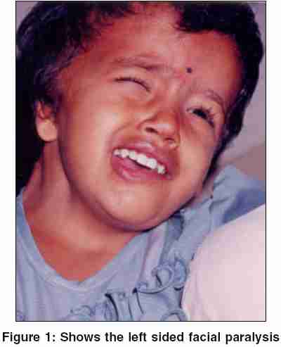
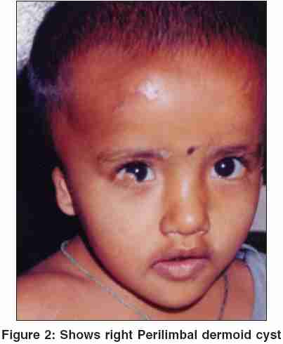
Indian Journal of Human Genetics, Vol. 11, No. 3, September-December, 2005, pp. 156-158 Case Reports Facio-auricular vertebral syndrome-a case report Reddy MV. V., Reddy* PP, Usha Rani* P, Hema Bindu* L Department of E.N.T, Osmania Medical College, Hyderabad, *Department of Environmental Toxicology, Institute of Genetics and Hospital Code Number: hg05031 Abstract Facio Auricular Vertebral (FAV) or Goldenhar syndrome is a very rare kind of syndromic deafness and is inherited as autosomal dominant. A study was taken up to understand the prevalence of this syndrome in children below the age of 14 years with hearing loss. Out of 1073 children with hearing impairment, Goldenhar syndrome was observed only in 1 (0.09%) case. The child suffered severe hearing loss. Facial paralysis and hemifacial microsomia were prominent features observed in the child. Facio-Auricular-Vertebral syndrome is therefore synonymously used with Goldenhar syndrome.Keywords: Hemifacial microsomia, Vertebral anomalies, Atresia, Dermoid cyst, Sensorineural, Hearing loss, Goldenhar syndrome Goldenhar syndrome is present in 1 in 1000 children with congenital deafness. In many cases, this syndrome goes unnoticed in Indian population due to the lack of knowledge about its features. The main aim of this study was to determine the incidence of this syndrome in Hyderabad population. FAV syndrome was first recorded by German physician Carl Ferdinand Von Arlt in 1845 and Goldenhar in 1952 defined the syndrome more clearly.[1] First and second branchial arch anomalies, otomandibular dysostosis and hemifacial microsomia was proposed as Goldenhar syndrome or facioauriculo vertebral syndrome.[1] This syndrome is also referred to as oculo-auriculo-vertebral (OAV) syndrome because of the association of ocular anomalies. The incidence of this syndrome is 1 in 3500 to 5000 live births and the male to female ratio of this syndrome is 3:2. The syndrome may or may not be associated with hearing loss. Conductive and / or sensorineural hearing loss is present in 50% of the patients with this syndrome. The etiology of the hearing loss is varied and may include missing or malformed outer ear (anotia and microtia), narrow or missing ear canals (atresia), abnormal skin cartilage on or in front of the ears (preauricular tags) and abnormalities in the middle or inner ear.[1] Goldenhar syndrome is mostly a sporadic condition and is inherited very rarely.[2] While many cases of Goldenhar syndrome associated with hearing handicap are reported from all over the globe, studies carried out from India are meager. Hence a study was taken up on children with congenital hearing loss to understand the prevalence of this syndrome in India. Case History The proposita is a three year old female child referred from an eye hospital for evaluation of hearing to the Govt ENT hospital. The proband is the second child of healthy non-consanguineous parents. The child was delivered vaginally at home and the birth weight of the child was 2000g (percentile). The child had birth asphyxia. Antenatal and intranatal periods were uneventful. Dismorphic features such as small mouth, the atretic right external ear canal and an accessory auricle (tag) in the left preauricular region were noted immediately in the child. Left facial paralysis and hemifacial microsomia was also observed [Figure - 1]. Micrognathia, limbal dermoid cyst was seen in the right eye [Figure - 2]. The first child, a boy and the third child, a girl were normal. Physical examination of the child showed undernourishment and weighed 10kgs. The child had short stature, frontal bossing, left facial paralysis (lower motor neuron lesion), short neck, bilateral microtia associated with meatal stenosis of the right ear and accessory auricle in the left preauricular region. X-ray of the chest showed revealed cardiomegaly, roentgenogram of cervical spine showed third hemivertebrae and fusion of fourth and fifth cervical vertebrae. No evidence of scoliosis. CT-scan of the temporal bone showed sclerosis of middle/inner ear bilaterally with hypoplastic left internal auditory canal and doubtful choleseatoma on right side with no evidence of bony erosion. Audiogram report showed bilateral severe sensorineural hearing loss in both the ears. Systemic examination did not reveal any cardiovascular or renal abnormality. Hemogram, complete urine examination, blood urea and serum creatinine were in normal levels. Cranial neurosonogram performed at the age of one month showed increased perventricular echogenicity, mild dilation of lateral ventricules associated with shaggy bulky outline of choroid plexus. Ultrasound findings of the child showed contracted gall bladder. Discussion The combination of several features such as microtia, hemifacial microsomia, lateral facial cleft, epibulbar dermoid and upper eye lid coloboma is described as oculo-auriculo-vertebral spectrum (OAV).[1] The presence of otic hypoplasia, lateral facial cleft and vertebral anomalies are the minimum criteria for the diagnosis of Goldenhar syndrome. The incidence of Goldenhar syndrome is extremely rare.[3] The etiology is not known, but is thought to be due to exposure to viruses or chemicals during pregnancy, due to abnormal vascular supply to the first arch and abnormality of mesoblastic development affecting the formation of vertebral and branchial systems.[4] Some researchers suggest that the disorder may be due to interaction of many genes with environmental factors (multifactorial inheritance). [4],[5],[6] Approximately 5000 children are born every year in United States with significant hearing impairment.[1] Goldenhar syndrome associated with deafness is rare and in most cases it occurs randomly without any apparent cause. Most cases are sporadic, but family history was also observed in certain cases. Individuals affected in successive generations have been observed.[7],[8] The ear anomalies include external ear tags and aural fistulae. [9],[10],[11],[12],[13],[14]In this case, the child showed malformation of the pinna associated with meatal atresia and auricular tags bilaterally. The behavior observation audiometry (BOA) showed 80-90 decibel hearing loss in both the ears. A 9 year old girl was reported with facial asymmetry, right ear with 2nd degree dysplasy and 3rd degree in the left ear[15] and a 10 day old patient was also reported with atresia and malformation of the tympanic cavity and ossicles.[16] With Goldenhar, the vertebral malformations include fusion or missing of the vertebrae. However in the present case the child showed the fusion of fourth and fifth cervical vertebrae. A small percentage also has mental retardation associated with this syndrome. Epibulbar dermoids and lipodermoids, coloboma of the eyelid, microphthalmia, strabismus and retinal anomalies are the associated ocular problems in Goldenhar syndrome.[17] Micrognathia limbal dermoid cyst was seen in the right eye of the child in this case. Children with FAV syndrome should be assessed for both vision and hearing. They may be subjected to numerous surgeries to correct the jaw and dental abnormalities. The structural anomalies of the eyes and ears in Goldenhar syndrome can be corrected by plastic surgery.[16] Expression of MSX class genes permit clear understanding of the variability and different degrees of severity of the anomalies of OAV spectrum. The MSX homeobox genes also play a crucial role in the differentiation of first branchial arch.[17] Acknowledgements We are greatful to the superintendent of Government ENT hospital and Principals of various schools for deaf for their kind co-operation. We are also highly indebted to the parents of the child without whose support the study wouldn′t have been possible.References
Copyright 2005 - Indian Journal of Human Genetics The following images related to this document are available:Photo images[hg05031f1.jpg] [hg05031f2.jpg] |
| |||||||||
