 |
| About Bioline | All Journals | Testimonials | Membership | News |
|
||||||
|
||||||
Indian Journal of Human Genetics, Vol. 18, No. 1, January-April, 2012, pp. 83-86 In silico prediction of exposure amino acid sequences of outer inflammatory protein A of Helicobacter pylori for surface display on Eschierchia coli Omid Teymournejad1, Ashraf M Mobarez1, Zuhair M Hassan2, Seyed M Moazzeni2, Bagher Yakhchali3, Vajihe Eskandari3 1 Department of Bacteriology, Tarbiat Modares University, Tehran, Iran Code Number: hg12014 DOI: 10.4103/0971-6866.96659 Abstract Background: Outer inflammatory protein A (OipA) is an outer membrane protein of Helicobacter pylori that is involved in inducing IL-8 and intracellular signaling. In this study, we have predicted exposure amino acid sequences of OipA for insertion in permissive sites of CstH subunit of Eschierchia coli CS3 pilli for bacterial surface display.
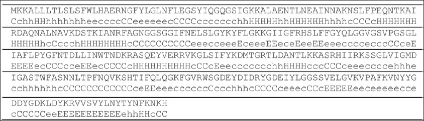
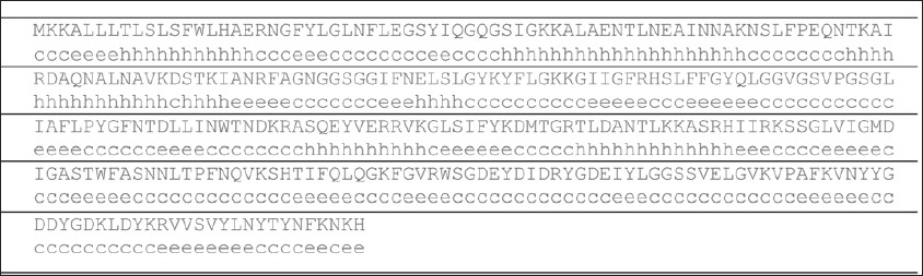
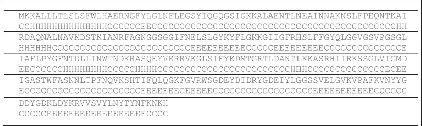
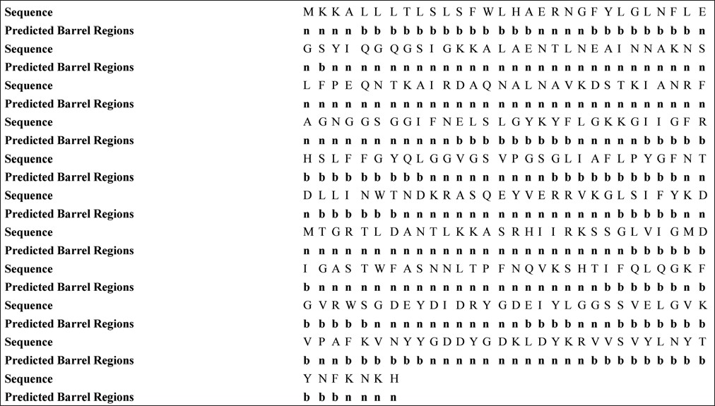
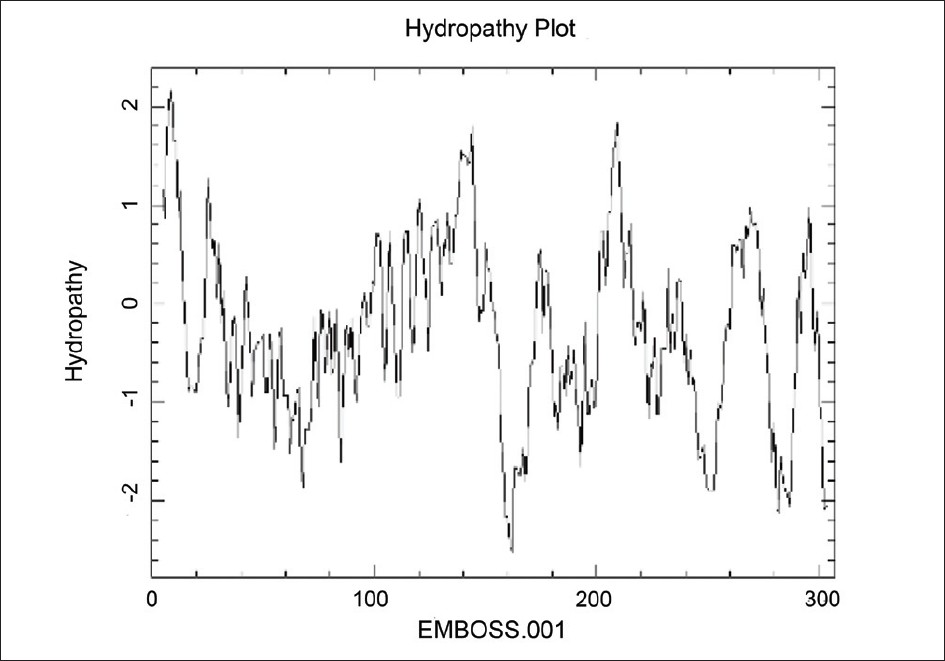
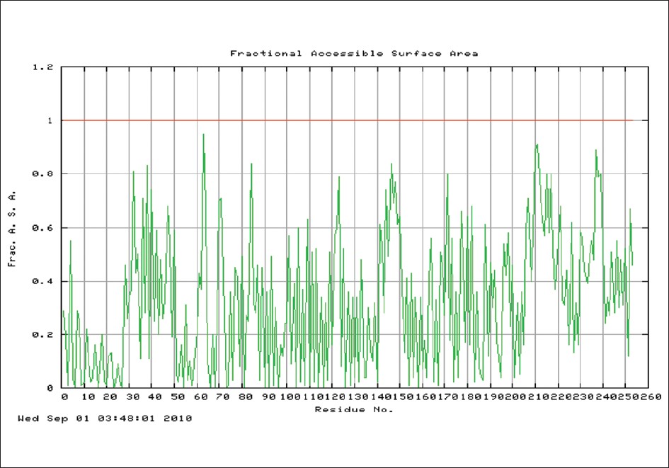
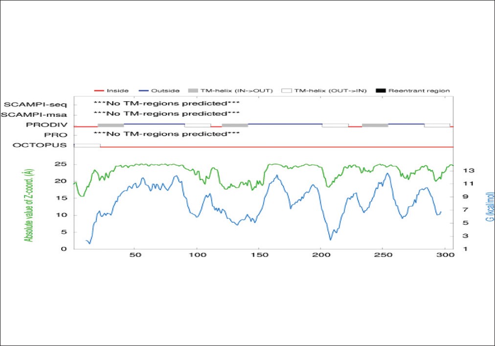
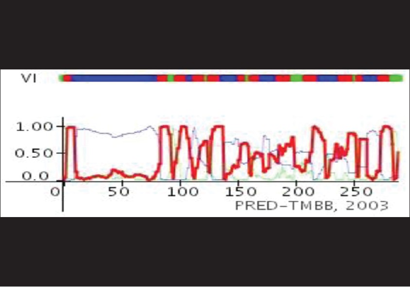
Keywords: CstH, in silico prediction, OipA, surface sequences Introduction Helicobacter pylori is a gram negative spiral bacterium that colonizes stomach for long time, and this colonization can lead to gastritis, peptic ulcer, and gastric cancer. [1] World Health Organization and International Agency for Research on Cancer have classified this bacterium as type one carcinogen for gastric cancer. [2],[3] H. pylori colonizes half of the world population and this colonization reaches up to 90% in developing country. [4] Outer membrane proteins have pivotal role in pathogenesis of this bacterium. [5] Most of these outer membrane proteins increase attachment to gastric epithelial cell and some of them induce host inflammatory responses. [5] Outer inflammatory protein A (OipA) is an outer membrane protein that is involved to increase pathogenesis of H. pylori. [6] Presence of this protein is linked to proinflammatory signaling of gastric epithelial cell, duodenal ulceration, gastric cancer, increase of H. pylori density, and neutrophil infiltration. [6],[7] OipA mutant of H. pylori revealed that this outer membrane protein associated with bacterial adhesion to gastric epithelial cell. Inducing IL-8 secretion by this protein is controversial; some reports showed that oipA mutants had not reduced IL-8 secretion but the others have reported about main role of OipA in induction of IL-8. [5],[6],[8] The aim of this research is computational studies for prediction of OipA exposure sequences of amino acid. Results of this study apply for insert in permissive sites of CstH subunit of Eschierichia coli CS3 pilli for bacterial surface display of outer inflammatory protein A on E. coli for detection adhesion motif and role of OipA in induction of IL-8 and proinflammatory signaling. Materials and Methods Databases: Amino acid sequences of OipA and CstH were retrieved from National Center for Biotechnology Institute and Uni-prot KB/Swiss-Prot. Accession number of CstH in NCBI and Uni-prot KB/Swiss-Prot were X16944 and P15488. [9],[10] Tertiary structure of CstH presents in Protein Data Bank. [11] Servers: Secondary structure of proteins predicted by PHD, SABLE, and GOR 4 servers. [12],[13] SignalP3.0 server detected signal sequence of OipA and CstH. [14] TBBpred, PRODIV-TMHMM, and TMRPres2D servers used for transmembrane topology prediction in bilayer lipid and external loops of OipA. CPH Models and PHYRE servers applied for finding of homology modeling and tertiary structure prediction. [15],[16],[17],[18] GETAREA and VADAR servers applied for prediction of accessible surface area amino acids among sequences of external loops. Pep state and pep window used for probability of expression in inclusion bodies and hydropathy plot of two proteins. Software: Swiss PDB viewer and Discovery studio applied for data analysis. [19],[20] Results In silico prediction of exposure amino acid sequences of OipA led to detection of six sequences of amino acid; 76-87, 106-112, 170-182, 222-230, 242-258, and 278-290. These sequences inserted between amino acid sequences 66-67, 100-101 and 109-110 of CstH were predicted by Eskandari et al. as permissive sites of CstH. Discussion Protein engineering is a branch of bioinformatics that is useful for progress of biological research. [21] In this study, we used from some tools of protein engineering for process of outer inflammatory protein A and prediction of exposure amino acid sequences for detection of adhesion motifs and sequences involve to proinflammatory signaling after bacterial surface display on E. coli. Carrier protein for OipA on surface of E. coli is CstH subunit of CS3 pili that previously was analyzed by Eskandari et al. for permissive sites. Prediction for secondary, tertiary structure, and hydropathy plot showed three sites for insertion of our sequences. [21] We applied three servers for prediction of secondary structure and compared these results with together [Figure - 1], [Figure - 2] and [Figure - 3]. SignalP3.0 server predicted seventeen N-terminal amino acids of OipA which do not present in mature OipA on the cell surface and this sequence was deleted before further analysis by other tools [Figure - 4]. Secondary structure of transmembrane proteins in prokaryotes has a little difference from eukaryotes. In outer membrane of prokaryotes, hydrophobic beta barrel integrate in bilayer lipid and famous tool for prediction of transmembrane topology is TBBpred server [Figure - 5]. [22] Tertiary structure of protein causes that some sequences of external loops get surface and the others get inside of protein. [22] Next step was tertiary structure prediction of OipA and detection of exposure amino acid sequences in external loops [Figure - 6], [Figure - 7] and [Figure - 8]. The CPH model server revealed that tertiary structure of OipA, same as VacA, was most likely an auto display protein and its insertion in the outer membrane was carried out by type V secretion system (T5SS). N-terminal long hydrophilic region confirms this finding [Figure - 6], [Figure - 7], [Figure - 8], [Figure - 9] and [Figure - 10]. Conclusion Exposure amino acid sequences of OipA and other outer membrane proteins have important role in interaction with stomach epithelial cell, and detection of these sequences is very useful for finding of receptors on surface of Gastric epithelial cell. [5] Receptor of OipA and adhesion motifs on this protein is unknown. Detection of exposure motifs aids to recognition of adhesion motifs and receptor of OipA on gastric epithelial cells. [23] In this study, we have predicted exposure amino acid sequences for insert to subunit CstH of CS3 pilli E. coli for surface display. References
Copyright 2012 - Indian Journal of Human Genetics The following images related to this document are available:Photo images[hg12014f5.jpg] [hg12014f3.jpg] [hg12014f9.jpg] [hg12014f8.jpg] [hg12014f10.jpg] [hg12014f1.jpg] [hg12014f4.jpg] [hg12014f7.jpg] [hg12014f6.jpg] [hg12014f2.jpg] |
| |||||||||
