 |
| About Bioline | All Journals | Testimonials | Membership | News |
|
||||||
|
||||||
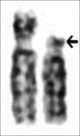
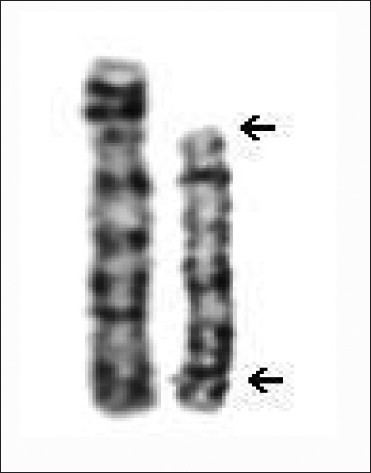
Indian Journal of Human Genetics, Vol. 18, No. 1, January-April, 2012, pp. 117-118 Wolf-Hirschhorn syndrome: A case demonstrated by a cytogenetic study Yamini S Pokale, Ajinkya M Jadhav, Ushang Kate Department of Cytogenetic, PreventiNe Life Care, 2nd Floor, RPT House, Plot No.6, Sector 24, Turbhe, Navi Mumbai-400705, India Code Number: hg12022 DOI: 10.4103/0971-6866.96677 Abstract We present a case with a 4p terminal deletion, evidenced in GTG-banded chromosome study. Phenotypic signs described in the classical Wolf-Hirschhorn syndrome were found on clinical examination of our patient. Keywords: Chromosomes, chromosome deletion, karyotyping, 4p, micrognathia, Wolf-Hirschhorn syndrome Introduction The Wolf-Hirschhorn syndrome (WHS) is a rare chromosomal disorder associated with a partial deletion of the short arm of chromosome 4. The syndrome is named after K. Hirschhorn and German U. Wolf who independently found the 4p-chromosome abnormality in the 1960s [1] and was first independently published in 1965 by Wolf et al. and Hirschhorn et al.[2]Wolf-Hirschhorn syndrome is a condition that affects many parts of the body. The major features of this disorder include a characteristic facial appearance such as a high forehead, highly arched eyebrows, epicanthal folds, fish-like mouth, low set ears, micrognathia, delayed growth and delayed developmental milestones, intellectual disability and seizures. It is characterized by congenital hypotonia, low birth weight, dental problems including missing teeth, and cleft lip or cleft palate. [3] Case Report The proband was the third child of non-consanguineous parents. The proband was born at term by normal delivery. He did not cry immediately after birth. Unfortunately proband died after 3 days of birth. He presented with distinctive facial features: he had low set ears, cleft palate (an opening in the roof of the mouth), and micrognathia (a small chin). Genitalia was observed male, but the infant had un-descended testes with no regular or well-developed scrotal sac. Examination shows foot deformity. [4],[5]Ultrasound examination of abdomen was reported to show a small kidney measuring 2 Χ 2 cm. According to Ballard gestational score, he corresponds to 32 weeks of gestation. Cytogenetic analysis from peripheral blood lymphocyte culture showed the proband having a terminal deletion of chromosome 4 at p14 region large enough to be detected using standard GTG-banding method. The karyotype revealed 46,XY,del(4)(p14)] [Figure - 1]. Discussion Wolf-Hirschhorn syndrome is a malformation syndrome associated with a terminal deletion of chromosome 4. This chromosomal change is also reported as 4p-. The size of the deletion varies among affected individuals; studies suggest that larger deletions tend to result in more severe intellectual disability and physical abnormalities than smaller deletions. WHS is a contiguous gene syndrome, in which the phenotype depends on the deletion of several different genes present in the homologous chromosome regions. Conventional G-banded cytogenetic studies detect a deletion in the distal portion of the short arm of one chromosome 4 involving band 4p14 in approximately 50%-60% of individuals with WHS. [6] The prevalence of WHS is estimated at approximately 1:50,000 births. Wolf-Hirschhorn syndrome is more common in females than in males, with a male-to-female ratio of 1:2. [2] Usually, the condition is detected in the newborn period because of dysmorphic features. The proband having a terminal 4p deletion large enough to be detected using standard GTG- banding method. Cytogenetic investigation of both the parents was done. Paternal karyotype was normal but maternal karyotype was found to be abnormal 46,XX,inv(4)(p14;q35) [Figure - 2]. Structural chromosomal rearrangements can have various reproductive outcomes ranging from infertility to a fetus with a normal karyotype or a fetus with unbalanced chromosomal rearrangements resulting in monosomy or trisomy of the these chromosomes or fetus can also inherit the apparently balanced rearranged chromosome of mother. [6] Structural chromosomal aberrations can lead to a wide variety of serious clinical manifestations, including mental retardation (MR) and congenital malformations. Chromosomal microarray technology as well as FISH (Fluorescence in situ hybridization) and/or G-banded cytogenetic studies may be necessary for complete characterization of the chromosome rearrangement associated with WHS. [6] The genetic counseling, prenatal diagnosis can be offered to determine the chromosomal status of subsequent pregnancies along with monitoring for fetal anomaly scan in all viable future pregnancy. References
Copyright 2012 - Indian Journal of Human Genetics The following images related to this document are available:Photo images[hg12022f2.jpg] [hg12022f1.jpg] |
| |||||||||
