 |
| About Bioline | All Journals | Testimonials | Membership | News |
|
||||||
|
||||||
Journal of Health, Population and Nutrition, Vol. 27, No. 5, Oct, 2009, pp. 612-618 Phenotypic Characterization of Salmonella Typhimurium Isolates from Food-animals and Abattoir Drains in Buea, Cameroon Jane-Francis T.K.Akoachere1, Nicoline F.Tanih1,2, Lucy M. Ndip1, and Roland N. Ndip1,2 1Department of Biochemistry and Microbiology, Faculty of Science, University of Buea, PO Box 63, Buea, Cameroon and
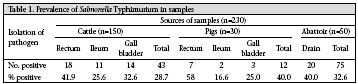
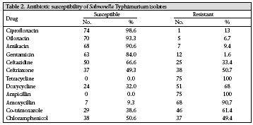
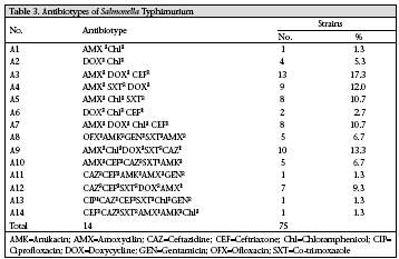
Code Number: hn09059 ABSTRACT Salmonella spp. have been extensively incriminated worldwide as common causes of bacterial gastroenteritis in humans, with food-animals serving as important reservoirs. The study was aimed at investigating cattle and pigs slaughtered in Buea as reservoirs of Salmonella Typhimurium and the susceptibility of isolates to antibiotics. In total, 230 specimens (comprising 50 each from the rectum, ileum, and gall bladder of cattle; and 10 each from same anatomical sites of pigs and 50 from abattoir drains) were analyzed for Salmonella using the standard microbiological, biochemical and serological techniques. Antibiotic susceptibility of the isolates was determined by the Kirby-Bauer disc-diffusion test. The isolates were characterized into biotypes using the API 20E kit, and results were analyzed using the chi-square test. Seventy-five (32.6%) of the 230 specimens were positive for S. Typhimurium, with pigs and abattoir drains presenting the highest level of isolation (40%). Biochemical typing grouped the isolates into five biotypes. Biotype I was the most prevalent (30.6%) while biotype IV was the least prevalent (9.3%) and was absent in samples from pigs. Antibiotic susceptibility studies revealed 14 antibiotypes based on antibiotics used in the study. The predominant antibiotype AMXR DOXRCEFR was recorded in 13 (17.3%) of the isolates. Multidrug resistance (to four or more antibiotics) was recorded in 50.7% (38/75) of the isolates. The most active drugs were ciprofloxacin (98.6%), ofloxacin (93.3%), amikacin (90.6%), and gentamicin (84%). All the isolates (100%) were resistant to tetracycline and ampicillin. Cattle and pigs were found to be reservoirs of S. Typhimurium in the environment of Buea, Cameroon, implying that foods from these sources, if not properly handled, could serve as vehicles for its transmission to humans. Key words:Antibiogram; Antibiotic resistance; Biotyping; Drug resistance, Microbial; Gastroenteritis; Salmonella infections; Salmonella Typhimurium; Cameroon INTRODUCTION Salmonella enterica , a Gram-negative, non-sporing, catalase-positive, oxidase-negative facultative anaerobic bacilli is a significant cause of morbidity and mortality in humans and animals, with multidrugresistant S. enterica serovar Typhimurium being an emerging problem (1-4). Contaminated food of animal origin, particularly meat products from cattle and pigs, is an important source of S. Typhimurium in human infections (5). S. Typhimurium has been described as a collection of variants that vary significantly in their host range and their degree of host adaptation (6). It is the third most common serovar causing human food-poisoning in some parts of the world. As pathogens, they have developed complex virulence mechanisms to evade host defence mechanisms (7). Although the organism does not cause clinical disease in pigs, subclinical infections constitute an important foodsafety problem throughout the world (8), and from a consumer viewpoint, continuing efforts are needed to reduce its occurrence in pork. S. Typhimurium can survive in the environment, and once established on a farm, contamination can be difficult to eradicate. It may spread from farm to farm through exchange of livestock, by wildlife, or in the runoff from fields and can disseminate into food-chains as a consequence of further cross-contamination at slaughter-houses. Due to the ability of Salmonellae to survive in meat and animal products that are not thoroughly cooked or not properly handled, animal products are the main vehicles of salmonellosis (4). Typing of S. Typhimurium provides information on strain diversity and improves the epidemiological analysis of outbreaks (9,10). Several methods, including biotyping, profiling of antibiotic susceptibility, phage-typing (11), pulsed-field gel electrophoresis (PFGE) (12), plasmid profile analysis (13), and various PCR-based techniques have been used for characterizing the organism. However, although phage-typing, PFGE, and PCR-based techniques have a high discriminatory power, they are more complex, cumbersome, and expensive, making these not suitable for routine investigation or for laboratories with limited resources as is common in developing countries. Zhou et al. employed biotyping as an effective means to the investigation and surveillance of S. Typhimurium-associated nosocomial infection (14); their findings demonstrated a link between infection in children and bacteria in hospital environments and carriers of medical staff. In the present study, we employed biotyping and antibiogram previously reported in our laboratory (15)as they offer advantagestosmallerlaboratories, such as ours, which are not optimally equipped. The emergence of multidrug-resistant (MDR) serotypes, especially S. Typhimurium definitive phagetype (DT) 104, has become a potential problem for animal husbandry and in human medicine (16-19). Animals infected with antibiotic-resistant Salmonella are an important source of resistance determinants that can transfer to human-infective Salmonella serovars. Considerable information abounds on distribution of serotypes and antimicrobial susceptibility of Salmonella of human and of food-animal origin in other parts of the world (8,20). However, a dearth of information exists on non-typhoidal Salmonella in most parts of Cameroon, including Buea (21). This study was, therefore, carried out to determine the role major food-animals (cattle and pigs) play as reservoirs of S. Typhimurium and also to study the susceptibility of isolates to antibiotics as drug resistance constitutes a serious health concern in this locality (15). This information will be important for obtaining epidemiological insight and for determining appropriate, empirical antimicrobial therapy in both human and veterinary medicines. MATERIALS AND METHODS Study design In total, 230 specimens were analyzed in the study. Fifty slaughtered cattle were sampled with 50 swabs each collected from the rectum, ileum, and contents of the gall bladder, giving 150 samples; 10 slaughtered pigs were sampled with three samples each collected from the same anatomical sites, such as the cattle for 30 samples; and 50 environmental samples were obtained from the abattoir drains. The samples were collected from different abattoirs, and sample sizes were selected based on convenience. More cattle were slaughtered than pigs in this and other abattoirs in Cameroon. The samples were collected during April-August 2006. Bacteriological analysis Specimens were collected and transported to the laboratory in selenite F medium following standard methods (22,23). They were incubated for 24-48 hours at 37 0C. The broth culture was aseptically streaked on Salmonella-Shigella agar (SS) and deoxycholate citrate agar (DCA) plates for the isolation of Salmonella. Plates were incubated at 37 0C for 18-48 hours, after which they were examined for colonies typical of Salmonella. Suspect colonies were streaked on nutrient agar plates to obtain pure cultures which were subjected to oxidase testing, gram-staining, and motility testing. Gram-negative short-motile rods and non-motile rods with characteristic red slope/yellow butt reaction on TSI either with the production of H2S or not were taken presumptively as Salmonella (22). They were further serotyped using agglutinating antisera (Murex Biotech Ltd., UK) based on the Kauffmann-White scheme as previously reported (24). Isolates were confirmed and classified into biotypes using the analytical profile index (API) 20E kit (Biomérieux SA, Marcy L’ Etiole, France) following the instructions of the manufacturer. Antibiotic susceptibility testing The Kirby-Bauer disc-diffusion test, which conforms to the recommended standard of the Clinical and Laboratory Standards Institute (CLSI), formerly National Committee for Clinical Laboratory Standards (NCCLS), was used as previously described (15,25). Briefly, a small inoculum of each pure bacterial isolate was emulsified in 3 mL of sterile normal saline in Bijou bottles, and the density was com-pared with a barium chloride standard (0.5 McFarland). A sterile cotton swab was dipped into the standardized suspension of bacterial cultures and used to evenly inoculate Mueller-Hinton plates (Biotec, England), and the plates were allowed to dry. Antibiotic discs with the following drug contents: gentamicin (GEN) (10 μg); amikacin (AMK) (10 μg); ciprofloxacin (CIP) (5 μg); ofloxacin (OFX) (10 μg); cefotaxime (CFT) (30 μg); ceftazidine (CEF) (30 μg); ampicillin (AMP) (10 μg); amoxicillin (AMX) (5 μg); tetracycline (TET) (30 μg); doxycycline (DOX) (10 μg); co-trimoxazole (SXT) (25 μg); chloramphenicol (Chl) (30 μg) (Antibiotic Becton, Dickson and Company, Sparks, USA; Le Point de Claix, France) were placed at least 15 mm apart and from the edge of the plates to prevent the overlapping of the inhibition zones. Plates were incubated at 37 0C for 24 hours, and the diameters of zones of inhibition were compared with recorded diameters of the control organism E. coli ATCC 25922 to determine the susceptibility or resistance of isolates to various drugs. These antibiotics were chosen based on the prescription practices for Salmonella in this locality and from the literature (16). Statistical analysis The chi-square test was employed to compare the prevalence in the different anatomical sites and biotypes. The differences were considered significant at p<0.05. RESULTS Prevalence of S.Typhimurium Of the 230 specimens analyzed, 75 (32.6%) were positive for S. Typhimurium. Samples from pigs and abattoir drains recorded a prevalence of 40% each while 28.7% was obtained from cattle (Table 1). The rectum had the highest isolation rate for cattle (41.9%) and pigs (58.3%) while the ileum had the least. There was, however, no significant difference (p>0.05) in the distribution of the organism in the different anatomic sites of animals. Characterization of isolates All the isolates were Gram-negative, motile, short rods which were oxidase-negative with a typical red slope/yellow butt reaction in triple sugar iron (alkaline slope/acid butt) with the production of high amounts of hydrogen sulphide and gas. These isolates were taken presumptively as S. enterica. Confirmation was based on API 20E reactions and polyvalent antisera. Biochemical characterization of the isolates resulted in five biotype patterns (I-V). Biotype I (30.7%) and II (26.7%) were the more prevalent while biotype IV (9.3%) was the least. An interesting finding was that biotype IV was isolated from cattle and abattoir drains but not from pigs. The major difference between biotype IV and other biotypes was the use of inositol, in which only biotype IV was observed to use this sugar. However, biotype I, III, and V were more frequently isolated from pigs compared to the other biotypes. There was no significant (p>0.05) difference in the distribution of biotypes. All biotypes from cattle also occurred in drains, indicating a probable link between biotypes from these sources. Antimicrobial susceptibility Susceptibility testing of isolates to 12 antimicrobial agents (Table 2) indicated that the quinolones (ciprofloxacin–98.6%; ofloxacin–93.3%) and the aminoglycosides (amikacin–90.6%; gentamycin–84%) were the most active drugs against the isolates. Tetracycline (100%) and ampicillin (100%), however, were the most resistant drugs. Marked resistance was also noted for amoxicillin (90.7%), doxycycline (68%), and co-trimoxazole (61.4%). Multidrug resistance was a common phenomenon observed with 38 (50.7%) of the 75 isolates. Fourteen antibiotic resistance patterns were obtained (Table 3). The predominant antibiotypes AMXRDOXRCE-FR (amoxycillin, doxycycline, ceftriaxone) and AMXRChlRDOXRSXTRCAZR (amoxycillin, chloramphenicol, doxycycline, co-trimoxazole, ceftazidine) were obtained from 13 (17.3%) and 10 (13.3%) of the isolates respectively. The least encountered patterns were AMXRChlR (amoxycillin, chloramphenicol), CAZRCEFRAMKRAMXRGENR (ceftazidine, ceftriaxone, amikacin, amoxycillin, gentamicin), CIPRCAZRCEFRSXTRChlRGENR (ciprofloxacin, ceftazidine, ceftriazone, co-trimoxazole, chloramphenicol, gentamicin), CEFRCAZRSXTRAMXRAM-KRChlR (ceftriaxone, ceftazidine, co-trimoxazole, amoxycillin, amikacin, chloramphenicol) as only one isolate each exhibited these patterns. DISCUSSION S. Typhimurium is a well-known zoonotic pathogen causing diarrhoea, pyrexia, and septicaemia in animals and humans. Non-typhoid Salmonella serovars remain a potential threat to human health, and beef cattle and broiler chickens are possible sources of these organisms in the environment (4). Although non-typhoidal salmonellosis in humans is usually a self-limiting disease confined to the intestinal tract, when infections spread beyond the intestine, or when immunocompromised persons are affected, it may have serious consequences requiring appropriate antimicrobial treatment. In animals, such symptoms can be lethal; so, prompt treatment with appropriate antimicrobial agents remains economically important. Hence, the surveillance of antimicrobial resistant strains is necessary for effective treatment and prediction of occurrence of resistant populations of prevailing biotypes. The public-health measures to reduce chances of infection, thus, take into consideration the presence of the organism in animals (26,27). This study was, therefore, conducted to determine the biotypes and antibiogram of S. Typhimurium isolated from slaughtered food-animals (cattle and pigs) in Buea, Cameroon, where no such data exist. S. Typhimurium was isolated from samples with a prevalence of 32.6%. The distribution pattern was similar in both species of animals sampled where the organism was more frequently isolated from the rectum (41.9% and 58.3% from cattle and pigs respectively) (Table 1). The ileum of both animals had the least occurrence of the organism. We associate our findings to the fact that these animals shed the organism in faeces when placed under stress during slaughter (28). Thus, rectal swabbing offers an easy method of surveying the carrier rate of a specific herd. There was, however, no significant difference (p>0.05) in the distribution of the organism in the anatomical sites of animals. The high prevalence of Salmonella in these animals could result from consumption of contaminated feed (29,30), or grazing plants that may have been contaminated through fertilization with untreated effluents or sludge. Classical biotyping characterizes strains by creating profiles for a set of biochemical tests. Previous studies employed biotyping as a marker for assessing the widespread outbreak of S. Typhimurium-associated infections (31,32). In our study, biotyping grouped isolates into five biotypes. Biotype I (30.6%) and II (26.7%) were the most frequently encountered. We observed a relationship between cattle and abattoir drains as all biotypes found in cattle were also obtained from drains. During slaughtering, the organisms are washed from cattle to open drains, and since the effluent is not properly disposed of, it may serve as a source for dissemination. Although we did not detect biotype IV in samples from porcine sources, we cannot declare the complete absence of this biotype from these sources as only a few swines were sampled due to a limited number of slaughtered swines in this locality and generally in abattoirs in Cameroon. In addition, although S. Typhimurium serotypes have been thought of as the prototypical broad-host-range serotypes, certain variants have been shown to have a narrow host range (6). We, therefore, speculate that the biotype IV could have a narrow host range. All other biotypes were present in all samples analyzed. Epidemiological surveillance of antimicrobial-resistant S. Typhimurium has become necessary for effective treatment and prediction of occurrence of resistant populations. Antibiogram of the isolates revealed marked susceptibility of isolates to quinolones—ciprofloxacin (98%) and ofloxacin (93.3%) (Table 2). Our results corroborate the findings of Esaki et al. (33) and Kawagoe et al. (20) who recently reported a marked susceptibility of S. Typhimurium to flouroquinolones that could be used in the treatment of infections caused by this organism. The high cost of these drugs in the study area discourages its over-use and may account for our observation of low antimicrobial resistance. Other active agents observed were the aminoglycosides—gentamicin (84%) and amikacin (90.6%). However, Kawagoe et al. recently reported resistance to these drugs in S. Typhimurium isolates from food-producing animals in Japan (20). We may not be certain as to these discrepancies but speculate that it may be related to differing prescription practices in these localities. It is noteworthy that all the isolates exhibited complete resistance (100%) to tetracycline and ampicillin. Esaki et al. reported complete resistance to ampicillin, dihydrostreptomycin, oxytetracycline, and chloramphenicol in DT104 and 104B S. Typhimurium (33). A marked resistance was also observed to doxycycline (68%) and amoxycillin (90.7%). Their use may result in treatment failure. Although we reported a resistance of 49.4% to chloramphenicol, its veterinary use for food-animals has been prohibited in some countries. Multidrug resistance was a common phenomenon in this study, being observed in 38 (50.7%) of the 75 isolates. Fourteen distinct resistance patterns (antibiotypes) were observed. Pattern AMXRDOXRCEFR was the most prevalent (17.3%) while CAZRCE-FRAMKRChlRGENR, CIPRCAZRCEFRSXTRChlRGENR, CEFRCAZRSXTROFXRAMKRChlR, and AMXRChlR were the least common (1.3%) (Table 3). The frequency of isolation of S. Typhimurium DT104 in food-animals worldwide has increased because of its spread and recent reports (20,34) on changes in resistance phenotype, or phage-type in MDR S. Typhimurium underscores the need for continuous monitoring of susceptibility pattern of S. Typhimurium from food-animals. Our findings of high levels of multidrug-resistant Salmonella in slaughtered cattle and pigs and in the environment highlight the potential risk of S. Typhimurium DT 104, with multidrug resistance becoming established in Cameroon. Thus, routine investigations at a national level for drug-resistant S. Typhimurium in food-animals and prudent use of antimicrobials remain a high priority. Frequent surveillance to track changes in the susceptibility pattern of the organism in the study area is, therefore, advocated. In conclusion, our results indicate that cattle and pigs could serve as reservoirs of S. Typhimurium in Buea, Cameroon and provide information on selection of antimicrobial therapy for infections due to S. Typhimurium in food-animals and for treatment of infections from these food sources. We advocate an urgent need for an organized Salmonella surveillance system that reports resistance patterns of S. enterica serotypes circulating in Cameroon. ACKNOWLEDGEMENTS The study was partly funded by the University of Buea, Cameroon. The authors are grateful for the collaboration received from the management of the Buea abattoir where samples were collected. Finally, the authors thank Mr. Monika Stephen for secretarial assistance. REFERENCES
Copyright © International Centre for Diarrhoeal Disease Research, Bangladesh The following images related to this document are available:Photo images[hn09059t3.jpg] [hn09059t2.jpg] [hn09059t1.jpg] |
| |||||||||
