 |
| About Bioline | All Journals | Testimonials | Membership | News |
|
||||||
|
||||||
African Health Sciences, Vol. 3, No. 2, August, 2003, pp. 68 - 76 Control of Taenia saginata by post-mortem examination of carcasses Wanzala W.a* , Onyango-Abuje J.A.b, Kang’ethe E.K.c, K. Hd Zessin, N. Mc,d Kyule, M.P.Od Baumann, Ochanda Ha, Harrison L.J.S.e a Division of Parasitology and Immunology, Department of Zoology, University of Nairobi, P.O. Box 30197,
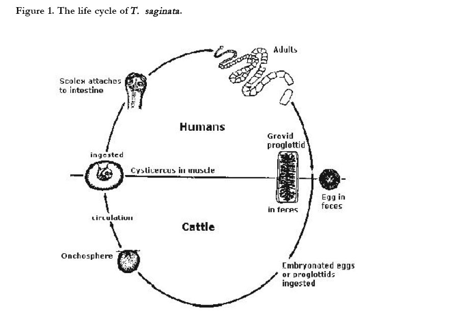
Nairobi, Kenya Code Number: hs03013 ABSTRACT Background: A study to curb transmission cycle of a zoonotic Taenia cestodiasis between humans and cattle is presented. Key words: meat inspection; post-mortem; zoonosis; bovine cysticercosis; taenia saginata INTRODUCTION Taenia saginata is a worldwide zoonotic cestode whose epidemiology is ethnically and culturally determined with estimates of approximately 50million cases of infestation worldwide with 50,000 people dying from this problem annually1 . Both adult and larvae forms hazardously affect health of their respective hosts, either directly or indirectly accompanied with severe secondary infections, particularly in human hosts2 . The occurrence of the larvae (Cysticercus bovis) in cattle musculature cause bovine cysticercosis while the adult worms in human small intestines cause taeniasis2 . In humans, the infestation is accompanied with mild symptoms ranging from nausea, abdominal discomfort, epigastric pain, diarrhea, vitamin deficiency, excessive appetite or loss of appetite, weakness and loss of weight to digestive disturbances and intestinal blockage2. However, in cattle, heavy infestation by the larvae may cause myocarditis or heart failure3 . The life cycle (Figure 1) and transmission of the parasite occur most commonly in environments characterized by poor sanitation, primitive livestock husbandry practices, and inadequate meat inspection, management and control policies4 . While ill-health caused by the adult worms in humans give rise to high medical costs5, the economic losses accruing from the condemned and downgraded carcasses due to treatment of carcasses before human consumption are substantial6,7. For instance, in Kenya, Botswana and Great Britain, such losses were estimated at, £1.0 million, £0.5 million8 and £1.2 million9 annually, respectively. For the African continent, an annual loss was reported to be US$ 1.8 billion4 under an overall infestation rate of 7%. In South America, where an overall infestation rate was estimated at 2.0%, bovine together with porcine cysticercosis caused an annual loss of US$ 428 million5. Various control measures in practice such as carcasses irradiation, public hygiene and education, mass chemotherapy (using praziquantel and/or albendazole and with praziquantel or niclosamide), cooking of meat at 57oC, deep freezing of meat at -10oC for 10 days, pickling meat in 25% salt solution for 5 days and buying only officially inspected meat10,2,11, have not been able to eliminate the parasite and even control it12. Other new potential approaches such as vaccination, chemotherapy and immunodiagnosis, have not been well developed for use and investigation still continues13,14,15,16,17. A promising Antigen-ELISA continues to be evaluated using diagnostic test evaluation tables17,18. Meat inspection method is therefore still the most important public health measure practised in controlling the transmission cycle of the parasite. The Kenya Meat Control Act19 regulations focus on the incision and examination for the presence of C. bovis only in the so-called predilection sites (cheek muscles of the head, tongue, heart and Triceps brachii). However, predilection sites, may not be the only sites in the carcass with the highest number of cysticerci as there could be great variations in terms of distribution of cysticerci between preferred sites20,21,22. Since meat inspection method is still the method of choice deployed in the control of human taeniasis at all the slaughterhouses, it was very necessary to re-evaluate it so as to assess its state of reliability, and hence the current study. METHODS Collection of the parasite eggs Taenia saginata proglottids were collected from human excrement in Mathare Valley Slums of Nairobi, Kenya. The proglottids were collected in physiological saline (0.15M NaCl) containing 200 units/ml of Crystapen benzylpencillin (Glaxo Laboratories, U.K.); 0.2mg/ml Streptomycin Sulphate (Glaxo Lab., U.K.) as antibiotics, and 5ug/ml fungizone (Squibb and Sons, Inc., New Jersey), as a fungicidal drug. While in the laboratory at the National Veterinary Research Centre (NVRC), Muguga, Kenya, the eggs were teased from the proglottids and washed through a tier of three sieves with 250nm, 150nm and 30nm apertures, respectively. The 30nm aperture sieve retained the eggs, which were then transferred into a universal bottle containing physiological saline and the antibiotics and stored at 4oC until required. The viability of the eggs was tested7 before calves’ infestation and eggs counted using McMaster slide. Naturally infested cattle
Naturally infested Zebu herds were identified through animal husbandry history and bovine cysticercosis reports from the District Veterinary Officer in Samburu District, Kenya. Ninety-six cattle were bled for serum, and tested for C. bovis circulating antigens by a sandwich Antigen-ELISA (Ag-ELISA)7,12,17. Based on the Optical Density (OD) readings, 25 steers about 1½ years old were selected. Sixteen of these were strong seropositive while nine were strong seronegative. The cattle were faecal sampled for nematode and fluke infestations using modified McMaster egg counting and Boray sedimentation techniques, respectively and dewormed accordingly. After three months, the cattle were slaughtered and carcasses examined for C. bovis using meat inspection procedures19and total dissection (cutting the musculature into very thin and transparent slices for the sake of recovering the parasite’s larvae)21,22. Artificially infested calves
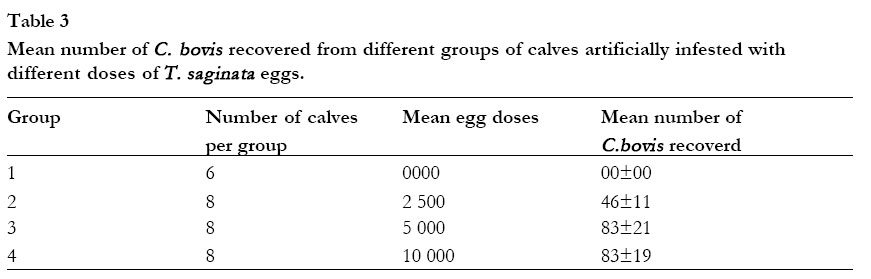
Thirty neonatal calves, 3 to 34 days old, were bought from a Ranch at Kapiti Plains, Machakos District. They were bled and the serum tested for circulating C. bovis antigens. The calves were kept worm/cysticercosis free in pens and fed on milk initially and later on calf weaner pellets and hay. Only one technician dressed in apron, gumboots and hand gloves to avoid man to animal infestation attended to them. The calves were divided into 4 groups (group 1(n=6) received sterile distilled water without any eggs (control), group 2 (n=8) received 2500 eggs each, group 3(n=8) received 5000 eggs each and group 4(n=8) received 10,000 eggs each) and infested when 2½ months old, after confirming their bovine cysticercosis-free state. Post - mortem examination
All experiments were performed in compliance with guidelines published by Kenya Veterinary Association (KVA) and Kenya Laboratory Animal Technician Association23. Meat inspection
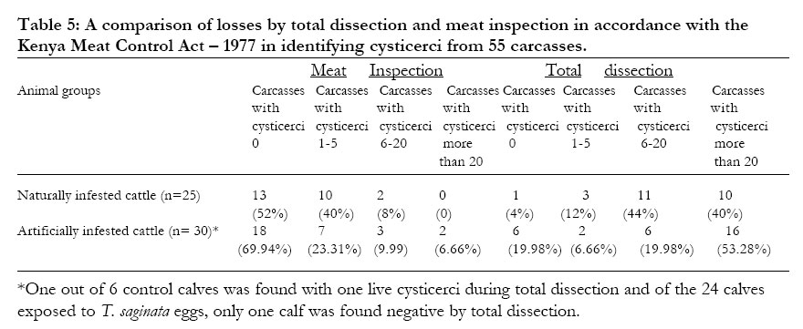
Meat inspection was done in accordance with the Kenya Meat Control Act19 which stipulates that the cheek muscles (masseter-external muscles and pterygoid-internal muscles of the head), tongue, heart and Triceps brachii must be deeply incised only once with a sharp knife and make three adjacent cuttings into the muscle to be examined for the presence of C. bovis before the blood covers the corresponding surfaces if at all the carcass was not well bled. For cheek muscles, two deep linear incisions were made parallel to the mandible from its upper muscular insertion. The tongue (also examined by palpation) was incised lengthwise on the lower surface from base to root while the heart was split from base to the apex and further incisions made into the muscles. Three deep, adjacent and parallel transverse incisions were made above the point of the elbow in the Triceps brachii. The Act further recommends that carcasses with 0 cysts should be passed on directly for human consumption, 1-5 cysts should be retained, frozen at -10oC for at least 10 days and released “unconditionally”, 6-20 cysts should be similarly treated as above but released conditionally to schools/institutions where proper cooking is expected to be done, over 20 cysts should be totally condemned. In rural areas where the disease is more prevalent and electricity is unavailable, the carcasses should be sliced and boiled for 2 hours at 77oC under the supervision of the inspecting officer. Total dissection
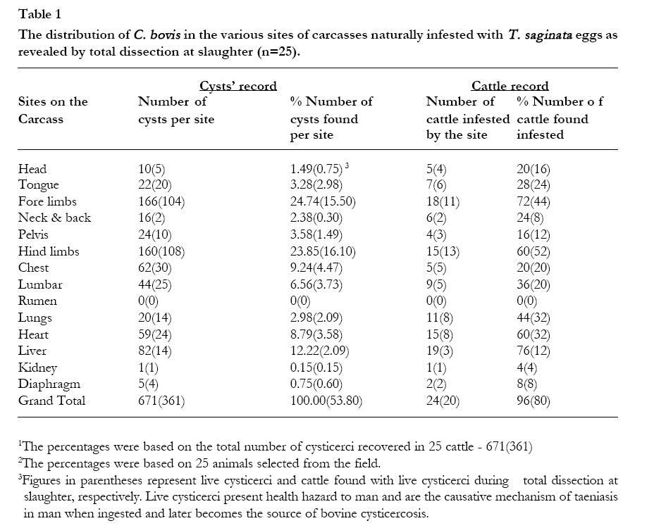
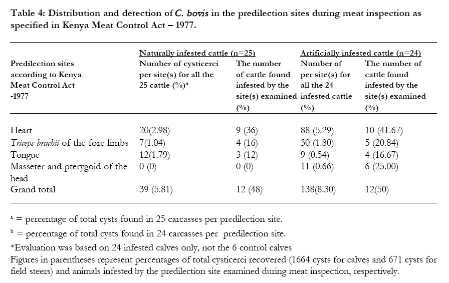
At slaughter the cattle were examined for cysticerci first by routine meat inspection procedures as stated above, followed by total dissection of the carcass. This was done by thinly slicing the entire musculature of the carcasses in order to recover the cysticerci. The number of cysticerci obtained in one half was noted and doubled to get the total number of cysticerci in the whole animal. The doubling method was adopted in accordance with statistically approved previous records based on long-term Standard Operating Procedures (SOP) set in NVRC laboratory, Muguga, Kenya on recovery of C. bovis from bovine carcasses7,21. However, visceral organs were not halved. In experimental calves, the whole carcass was examined for cysticerci because the cattle were small in size. Total dissection method was taken as a gold standard method of validity (a method that gives the true status of bovine cysticercosis infestation in cattle), against which post-mortem meat inspection method was compared. RESULTS Distribution of T. saginata cysticerci in carcasses of naturally infested cattle The distribution of cysticerci in various sites of the carcasses of 25 naturally infested cattle is shown in Table 1. All sites except the rumen had varying numbers of both live and dead cysticerci, with skeletal muscles carrying the highest (as high as 24.74%) while visceral organs carrying the least (as low as 0.15%). All the 25 animals were found infested (96%) except one. More live cysticerci (53.80%) were recovered in skeletal muscles than the dead ones (46.20%) in visceral organs (Table 1). By physical appearance, dead, degenerate or calcified cysticerci clearly formed identifiable spots of white and fibrotic lesions, while the viable cysticerci were pinkish-red in colour. However, cysticerci, which evinced true viability and could therefore be counted as live, evaginated upon staying in cattle bile overnight, while the dead ones remained intact albeit pinkish colouration. This therefore stopped further histopathological confirmation of dead cysticerci following their morphological confirmation of their originality and true identity from the voucher specimens stored in NVRC laboratory. Distribution of T. saginata cysticerci in carcasses of artificially infested calves The results of the distribution of cysticerci between parts of the carcasses of calves are shown in Table 2. All sites except the Kidneys had varying numbers of both live and dead cysticerci, with the visceral organs carrying the highest (as high as 22.00% for liver) while the skeletal muscles generally carrying the least (as low as 4.09% for Lumbar). All the 24 treated calves were found infested (95.83%) except one. Total dissection, like in natural infestations, revealed cysticerci in all the 30 experimental calves except in one of the 24 treated calves and one of the 6 control calves (Table 2 and 5). More live cysticerci (52.01%) were recovered in skeletal muscles than the dead ones (47.99%) in visceral organs (Table 2). However, egg doses administered to calves did not have influence on the recovery of cysticerci at autopsy (Table 3). Meat inspection findings in naturally and artificially infested cattle Meat inspection detected cysticerci in 12 out of 24 animals (50%) in artificially infested calves. Of these calves, 10 (41.67%) had cysticerci in the heart, 6(25.00%) in the cheek muscles (masseter-external muscles and Pterygoid-internal muscles), 5 (20.83%) in the Masculus triceps brachii (shoulder muscles) and 4 (16.67%) in the tongue (Table 4). The six control calves were not found with any cysticerci. All the predilection sites of the carcasses of artificially infested calves were found with cysticerci but most were found in the heart (5.29%), Masculus triceps brachii muscles (1.80%), masseter and pterygoid muscles (0.60%) and tongue (0.54%) in that order (Table 4). In animals with natural infestations, meat inspection revealed a prevalence rate of 48% (12 out of 25 animals) with a considerably varied detection rate of cysticerci in different carcasses and predilection sites (Table 4). Of these animals, 9 (36%) had cysticerci in the heart, 4 (16%) in the Masculus triceps brachii (shoulder muscles) and 3 (12%) in the tongue. In natural infestations, most cysticerci were found in the heart (2.98%), tongue (1.79%) and Masculus triceps brachii muscles (1.04%), in that order (Table 4). A comparative analysis of meat inspection and total dissection in detecting C. bovis in carcasses from economic loss and enhancement of transmission cycle point of views. Detection of C. bovis by the two methods (meat inspection and total dissection) was often in an inverse proportion (Table 5). From the results shown in Table 5, total dissection showed that a lot of infested animals are passed on for human consumption by meat inspection under the current Kenya Meat Control Act-1977, thus, enhancing a continued transmission cycle of the parasite between humans and cattle. By total dissection, 40% and 53.28% of the animals from natural and artificial infections, respectively, would be totally condemned, a great loss to the farmer and butcher business. While meat inspection method never condemned any animal from natural infections but only 6.66% from artificial infections, thus, allowing a lot of infested carcasses to enter the transmission cycle. DISCUSSION
In both naturally and artificially infested cattle, total dissection revealed cysticerci infestation in 24 out of 25 cattle selected from the field and 23 out of 24 calves exposed to the eggs of T. saginata, giving prevalence rates of 96 and 95.8%, respectively (Table 1 and 2). While the corresponding prevalence rates by meat inspection were 48 and 50%, respectively (Table 4). In these 47 infested carcasses, cysticerci were found distributed between and within carcasses’ sites in varying numbers and surprisingly, even more in sites not specified in the Act-1977 as predilection sites (Table 1 and 2). This, together with other factors (light intensity at the inspection points, room ventilation, nature of the knife in use, poor bleeding of the carcasses, uncoordinated training programmes of the officers, pressure of work, corruption and poor human eyesight) previously identified24,25 completely undermine the sensitivity of meat inspection. Crucial to this Act, is its limitations such as, fewer predilection sites and only a small area within a given predilection site being allowed to be incised and examined so as to avoid mutilation of the carcass. The results of the total dissection were in agreement with the previous studies 3,20,26,28, in terms of distribution of cysticerci in various parts of the carcasses and ranking of these parts, albeit great variations. The variations depend on a number of factors such as blood kinetics and animals’ daily activities3. Any geographical and environmental factors affecting blood kinetics in the animal affect the distribution of oncospheres as well and hence the predilection sites during meat inspection28. Total dissection results showed a high density of both live and dead cysticerci in the hind limbs, fore limbs, liver, chest, heart, lumbar, pelvis, tongue, lungs, neck and back, head and diaphragm in that order (Table 1 and 2). From these results, all the parts of various carcasses were equally important as predilection sites for cysticerci and could be equally used during routine meat inspection at slaughterhouses except for rumen, fat layers, spleen and skin (the last three sites not included in the results but during total dissection, they never had any cysticerci). However, of the parts examined, hind limbs, ribs, lungs, neck and back, liver, lumbar and pelvis, need to be considered by the Act-1977 and larger areas of these predilection sites be examined during routine meat inspection. The age-dependent immunity26 of an animal had an important role to play in fighting against infestation and re-infestation of cysticerci29. The re-stimulation of animals’ immunity following continuous invasion of oncospheres, would explain the development of a strong immunity which did not allow further development of more cysticerci from invading oncospheres but spared viable cysticerci from the initial infestation26,30,31,32. This phenomenon further helps to explain the occurrence of both live and dead cysticerci and their corresponding variations in predilection sites examined (Table 1 and 2). In artificial infestations, the intensity of cysticerci was highest in the visceral organs (liver - 22.00% and heart - 17.13%) followed by the limbs (hind limbs - 15.02%, fore limbs 13.35% and chest - 6.49%) (Table 2) This pattern reversed in naturally infested cattle with the limbs (fore limbs 24.74%, hind limbs - 23.85% and chest - 9.24%) harbouring the highest number of cysticerci followed by visceral organs (liver - 12.22% and heart - 8.79%) (Table 1). The explanation for this differential location of cysticerci lies in the animals’ daily activities and blood kinetics28, which determines the distribution of cysticerci within the animal. These results indicated that in either group of cattle, total dissection was, in practice, twice as efficient in the detection of bovine cysticercosis as routinely used meat inspection (Table 1, 2 and 4). While Table 5 shows the insensitivity of meat inspection method. Though robust, total dissection cannot however, be used in slaughterhouses as meat inspection method because it is such a tedious, timeconsuming method and greatly lowers the quality of meat and its marketability. The routinely used meat inspection method is disadvantaged by the fact that it is directly dependent on human judgement by physical observation, identification and removal from specified distinct predilection sites only3,20. This therefore means that an animal could be diagnosed negative even if cysticerci were located elsewhere in the carcass being examined. During the inspection of various carcasses, it was realised that except for the dead, degenerate or calcified cysticerci which often formed spots of white and fibrotic lesions, a careless meat inspector will most likely miss out quite a number of viable cysticerci which blend with the pinkish-red colour of the meat and which are then passed on for human consumption. This significantly lowers the sensitivity of the routinely used meat inspection method. This unreliability and low detection rate has previously been observed especially in lightly infested cattle6,25. While the implementation of better and novel diagnostic methods for bovine cysticercosis remains at the trial stages16,33,34,35,36, the advocates25 of meat inspection method should know that it is still an insensitive method to be used for the control of human taeniasis and bovine cysticercosis. This is clearly shown in Table 5 where a comparative analysis of meat inspection and total dissection in detecting C. bovis in carcasses from economic loss and enhancement of transmission cycle point of views was critically evaluated. To effectively improve meat inspection procedures, there is need therefore to increase the area and number of predilection sites observed during inspection and vary them according to the nature of the animals, their husbandry history and the target human population for consumption. In addition, develop new control approaches such as vaccination, chemotherapy, health education, improved sanitation, favourable socioeconomic conditions and immunodiagnosis to complement meat inspection procedures. ACKNOWLEDGEMENTS
The authors wish to thank Dr. L. Wamae for sampling, buying and transporting cattle to National Veterinary Research Centre (NVRC) Muguga, members of staff of Helminthology Division, NVRC, for their technical assistance. This work was supported by a financial grant from European Union (EU). The paper is published with the kind permission of the director of Kenya Agricultural Research Institute (KARI), Kenya. Besides, l wish to acknowledge the constructive suggestions and criticisms received from the two reviewers appointed by the Editor, Journal of African Health Sciences. To all, we are very grateful to their support and permission. REFERENCES
Copyright © 2003 - Makerere Medical School, Uganda The following images related to this document are available:Photo images[hs03013t4.jpg] [hs03013f1.jpg] [hs03013t5.jpg] [hs03013t1.jpg] [hs03013t3.jpg] [hs03013t2.jpg] |
| |||||||||
