 |
| About Bioline | All Journals | Testimonials | Membership | News |
|
||||||
|
||||||
African Health Sciences, Vol. 6, No. 2, June, 2006, pp. 81-85 Antifertility effect of aqueous and ethanol extracts of the leaves and roots of Asparagus africanus in rats Geremew Tafesse1, Yalemtsehay Mekonnen2, Eyasu Makonnen3
Corresponding author, Dr. Yalemtsehay Mekonnen, Department of Biology, Faculty of Science, Addis Ababa University, P. O. Box 1176, Addis Ababa, Ethiopia yalemtsehay@yahoo.com, yalemt@bio.aau.edu.et Fax: 00251-11-2755296 Tel: 00251-11-2763091 Code Number: hs06019 Abstract Background: Asparagus africanus is claimed to have use in reproductive
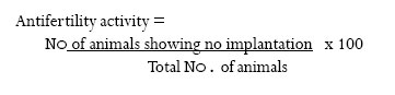
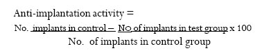
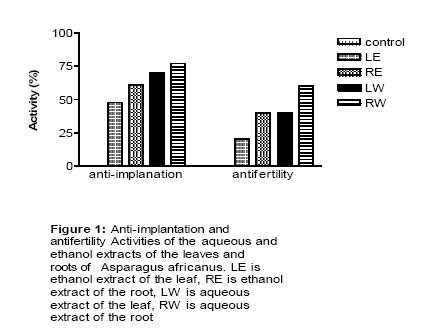
related health problems in some areas of Ethiopia. Key words: Antifertility,Anti-implantation, Asparagus africanus,aqueous extract, ethanol extract, leaves, roots, rats Introduction Asparagus africanus (L) is one among the many plants with traditionally claimed antifertility properties in Ethiopia. This plant belongs to the Family Liliaceae. It is a perennial climbing or erect shrub that can grow between 700 and 3800 m above sea level. However, it is widely distributed and suitably grows higher up to 6m at the altitude range of 1450 and 2900 m 1 . Its vernacular name is “Saritti”; this plant has been known for a long time to serve as a traditional cure for problems related to reproductive health. An infusion of its roots is employed as a remedy for venereal diseases. Moreover, at parturition the infusion of the root being mixed with water is taken by women to facilitate the process2, 3 . In rural areas of Bale zone (southern parts of Ethiopia) the leaf or root of Asparagus spp. is traditionally chewed to facilitate childbirth4 . Steroidal saponins, the most probable component of estrogen, were isolated from its roots5 . The steroidal saponins, which were isolated from the roots of its related species of A. officinalis, were reported to have uterine contractile property6 . The objective of this study is, therefore, to investigate the possible antifertility effect of the water and ethanol extracts of the leaves and roots of A. africanus to substantiate its use as herbal medicine. Materials and methods Plant collection and extract preparation The leaves and roots of Asparagus africanus were collected around Meki town about 130 km and the shore of Lake Langano about 210 km from Addis Ababa, the capital city of Ethiopia in May 2001. A botanist identified the plant and sample specimen was kept in the National Herbarium of Addis Ababa University under voucher number 1/2001. The leaves and roots were chopped with knife, and made dry in shade independently and then ground into powder with mortal and pestle. Measured amounts of the powder were macerated in distilled water (1 g / 6 ml, w/v) and in 90% ethanol (1 g / 4 ml, w/v). The macerated extracts were stirred with magnetic stirrer for 24 hrs at room temperature. Each extract was then flittered using cotton and Watman filter paper No1 7. After filtration, the water extracts were lyophilized with a lyophilizer; while ethanol was evaporated from the ethanol extracts using Rota vapour at 40 ° C 8 . The resulting partially solid extracts were stored at -20° C until used. Experimental animals Male and female albino rats were obtained from Faculty of Medicine, Addis Ababa University and let to breed continuously in the animal house of the Department of Biology,Addis Ababa University by allowing to be paired for any length of time as needed for mating.They were kept in cages in animal house with a 12:12 hr light-dark cycle. All the conditions were the same to all animals. They all made to feed on pellet diet and water ad labium. The newly born female rats were separated from males at the age of four weeks in order to prevent uncontrolled mating. Virgin female rats 10 to 11 weeks of age weighing between 195 and 200 g were used for both in vivo and in vitro experiments. In vivo test There were four experimental groups, i.e., aqueous extract of the leaf (LW), aqueous extract of the root (RW), ethanol extract of the leaf (LE) and ethanol extract of the root (RE).There was also one control group for the water and ethanol extracts. Each rat both in the experimental and control groups was kept singly in a cage to acclimatize for a week9. After a week of acclimatization experimental groups were administered with 300mg/ kg body weight of the extract by gavages, while the control groups received same volume of the vehicles. Each extract and the vehicle were dissolved in a 0.5 ml solvent. Distilled water was used to dissolve the aqueous extracts, while 70% ethanol and distilled water in the ratio of 2: 3 were used to dissolve the ethanol extracts. While dosing continued, males of proven breeding ability were introduced into each cage on the ninth day according to the method described by Williamson et al.9 . Vaginal lavages were used to determine the presence of sperm every morning. The animals were separated immediately after confirming mating. The oestrous cycle was monitored and the animals were still cycling after the nine days of dosing. By the method described by Williamson et al.9 laparatomy was performed on day 20 of pregnancy.The animals were sacrificed, and the number of implantation sites was determined. Weight gained by each rat of all the groups was recorded. Anti-implantation and antifertility activities were expressed as percentages using the following formula (9):
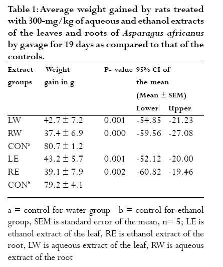
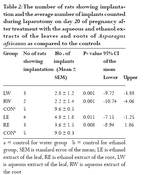
In vitro test A gentle blow on the head killed each rat.The abdomen was opened, and the uterine horns were cut at their junctions with the fallopian tubes, and placed in a dish containing De Jalon´s solution. For each experiment a uterine strip was set up in a thermostatically regulated organ bath that contains the solution, which was maintained at about 370 C and gassed with air8, 9. The uterine strip was tied with a string to a transducer (Grass FT.03) that was connected to Grass Polygraph model 07 to record contractions. A tension of 1gm was applied to the tissue and was allowed to equilibrate for at least 30 min before starting the test. Acetylcholine (Ach) was used as a stimulant to record contractions. A dose response curve was plotted with acetylcholine at a final bath concentration each of 40ng/ ml, 80ng/ml, 160ng/ml and 320ng/ml. Each time, the added acetylcholine was left in contact with the tissue for 30 seconds and then washed with De Jalon’s solution. It was then left to resume its normal contraction. Acetylcholine (80ng/ml) was selected as the control concentration that induced sub-maximal contraction of the uterine tissue. A given concentration of an extract was added in the organ bath and left in contact with the tissue for 5min.Acetylcholine (80ng/ml) was added at the end of the 5 min in the presence of the extract and then washed after 30 sec. After rhythmic contraction resumed, the same concentration of acetylcholine was added in order to establish the reversible contractile capacity of the tissue and test the extent the extract acted upon the uterine tissue.The same procedure was repeated whenever different extracts at different concentrations were tested. Each extract was tested at 80µg/ml, 160µg/ml, 240µg/ml and 320µg/ml, as final bath concentration. Contraction peaks recorded by the polygraph were measured in cm.The contraction peak of the control acetylcholine (80ng/ml) was taken as a reference (100%). The peak produced by 80ng/ml of acetylcholine was compared with that produced by the extract plus acetylcholine. The effect of the extract at a given bath concentration was then recorded by measuring the length of the peak produced by the combination of the extract and acetylcholine. Each value was converted to a percentage contraction by considering a 100% acetylcholine contraction.Then the mean percentage contraction was taken to compare the result10. Seven uterine strip preparations were used for each sample tested. Statistical analysis For the in vivo anti-implantation tests the mean ± SEM weight gain and the mean ± SEM number of implants in each test group were compared with the respective control groups. Independent Student’s t-test was used to analyze the result. In the in vitro test, the mean (± SEM) percentage tissue contraction due to acetylcholine was also compared with the mean (± SE) percentage tissue contraction due to acetylcholine with the extract at each bath concentration. One-way ANOVA test was used to analyze the results. In each test a 95% confident interval was used. Results In vivo test The weight gained with all extracts was significantly less than that of the controls (p < 0.005) as shown in table 1. The weight gained by the aqueous extracts of the leaves and roots of the plant was not significantly different from that gained by their ethanol counterparts. The root extracts, however, appeared to show less weight gain than the leaf extracts. But the difference is not significant.All rats in both control groups have shown implantation, while some rats among those, which received aqueous and ethanol extracts of both parts of the plant have shown no implantation (Table 2 ).The average numbers of implants in the pregnant rats with both the aqueous and ethanol extracts of the leaves and the roots of the plant were significantly less than that of the pregnant rats which received the vehicle (p < 0.005). Though it appeared that the root showed less number of implants than the leaf, and the aqueous extract showed less number of implants than the ethanol extract, the differences were not significant. The anti-fertility and anti-implantation activities produced by the aqueous extract of the leaves were 40% and 70%, respectively, while those of ethanol extract were 20% and 48%, respectively (Figure 1). The same figure shows that the anti-fertility and anti-implantation activities produced by the aqueous extract of the roots were 60% and 77%, respectively, while those of ethanol extract were 40% and 61%, respectively. Figure 1 also shows that the root showed more antifertility and anti-implantation activities than the leaf, and the water extracts were more potent than the ethanol extracts. In vitro test As shown in Figure 2, all extracts except the ethanol extract of the leaves increased the uterine contractile activity of acetylcholine significantly (p < 0.05). Furthermore, the capacity of the extracts to increase uterine contractile activity of acetylcholine was concentration dependent (Figure 2).The aqueous extracts of both the leaves and roots showed significant increase (p < 0.05) in the contractile activity of acetylcholine as compared to the ethanol extract (Figure 2).The extracts alone did not effect contraction. The root extracts were significantly more potent (p < 0.05) than the leaf extracts as depicted in the same figure. Discussion The less weight gain in the test groups as compared to the control groups could indicate that there were few implantations with all the extracts. The number of rats that had shown no implantation and the reduction in the number of implants suggests the possible antifertility activity of the extracts.The anti-fertility activity observed by the aqueous extract of the root was more or less similar with the result reported by Woo et al. 11 for Dectamus albus. According to Woo et al. 11 the anti-fertility activity of the methanol extract of the root bark of this plant was 60%. The reduced anti-fertility activity observed by the ethanol extracts in the present study might be due to the presence of more amount and/or number of active principles and/or better efficacious active principles in the aqueous extract than the ethanol extract. Steroidal saponins, one of the active principles of most anti-fertility agents12, 13 were reported earlier to exist in the root of A. africanus 5.The antifertility and antiimplantation activities observed in the present study might be attributed to these active principles. The ability of all the extracts to potentiate acetylcholine effect on uterine contraction in the present study suggests the possibility of interaction of the plant material with endogenous acetylcholine to induce abortifacient effect.The results obtained in the present study correlate with that reported earlier by Makonnen et al (14) for Ricinus communis and Jatropha curcas seeds on guinea pig uterus and by Mekonnen (10) for Moringa stenopetala on mouse uterus. The fact that the root was found to be more potent than the leaf in potentiating the acetylcholine effect suggests that more amount and/or number of active principles and/or active principles with better efficacy might be present in the root than in the leaf. The leaf and root of this plant may possess hormonal properties that can modulate the reproductive function of the experimental rats. Different mechanisms might have been involved to produce the antifertility activity that needs further elucidation. The present study demonstrated the possible therapeutic application of the leaf and root of A. africanus in fertility control. However, further study on the pharmacokinetic profile, safety and active principles of the plant have to be carried out. References
Copyright © 2006 - Makerere Medical School, Uganda The following images related to this document are available:Photo images[hs06019t2.jpg] [hs06019f2.jpg] [hs06019f1.jpg] [hs06019t1.jpg] |
| |||||||||
