 |
| About Bioline | All Journals | Testimonials | Membership | News |
|
||||||
|
||||||
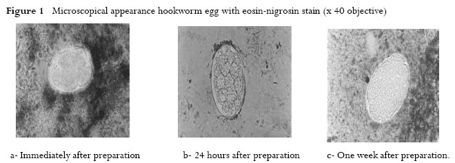
African Health Sciences, Vol. 7, No. 1, March, 2007, pp. 33-36 Substitution of Malachite Green with Nigrosin - Eosin Yellow Stain in the Kato-Katz method: microscopical appearance of the helminth eggs Emmanuel I.Odongo-Aginya1; Narcis Kabatereine2; Siefert Ludwig3; HenryWabinga4; Alan Fenwick5; Antonio Montresor6. 1) Uganda Virus Research Institute P.O.Box 42 Entebbe Uganda (East Africa). Corresponding author Antonio Montresor World Health Organization 63 Tran Hung Dao Street Mail P.O. Box 52 Ha Noi - Vietnam Tel +(84 4) 943 3734 /5 /6 ext 29 Fax +(84 4) 943 3740 e-mail montresora@vtn.wpro.who.int Code Number: hs07007 Abstract Background:The Kato-Katz thick smear technique is the standard technique recommended by the World Health Organisation for the
quantitative diagnosis of Schistosoma mansoni and other intestinal helminth infections.The major problem of the technique is that a few
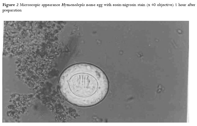
hours after the preparation of slides hookworm eggs over clear and disappear due glycerin. Keywords: Kato - Katz method, pictorial illustration, Odongo-aginya modification, helminth eggs, Uganda. Introduction The Kato-Katz thick smear technique is the standard technique recommended by World Health Organisation (WHO) for the quantitative diagnosis of Schistosoma mansoni and other intestinal helminth infections1. The glycerin, in the malachite green in Kato Katz’s technique, functions as a clearing agent while the malachite green besides being a dye, is bactericidal 2,3 . The Kato-Katz methods require between 1 to 2 hours before the glycerin clears the background of the stool smear on the slide for accurate visualization of most helminth eggs 1. The major problem of the technique is that few hours after the preparation of the slide hookworm eggs are difficult to recognize due to overclarification by glycerin 1. The aim of this paper is to show the appearance of the helminth eggs when malachite green is replaced with a stain comprised of nigrosin and eosin yellow in formalin4. Several field studies confirm the simplicity, quality, and cost effectiveness of the proposed modification5-9, a visual reference of the results of the method can be useful to facilitate the recognition of parasite eggs by microscopists willing to adopt this methodology. Material and Method The malachite green in the Kato-Katz technique is substituted by the following compound stain: 5% eosin yellow in 10% formalin, mixed 1:1 with 7.5% nigrosin in 10% formalin4. Cellophane cover slips cut in 25x40 mm pieces are soaked in 50% glycerin. Stool specimens are strained through a stainless steel sieve as used in Kato- Katz’s method. Through a template, providing 41.7 mg of feces, the strained stool is delivered on a microscope slide and a drop of the compound stain (about 10-50μl) is added to the stool on the slide and stirred using the corner of another slide. A cellophane cover slip presoaked in 50% glycerin is picked with a pair of forceps and excess glycerin on it is blotted out on an absorbent paper. The cellophane cover slip is then placed on the stained stool on the slide. The slide is inverted upside down and pressed down gently on tissue paper or any absorbent paper to spread out the stool smeared on the slide and to remove excess of the stain from the slide. A well-prepared slide has a pinkish background and is thin enough to read the face of a wristwatch through it. Photographs of the helminth eggs are presented in figure 1, 2 and 3. The photographs were taken using an Olympus Photomicroscope B 20 from slides prepared in the Department of Pathology Makerere Medical School according to Kato-Katz and Odongo-Aginya’s methods. The stool specimens were collected from Kigungu residents as a part of Odongo-Aginya, Doctor of Philosophy (Ph.D) study. Kigungu is a fishing village on Lake Victoria shoreline, Entebbe Uganda. Photographs were taken immediately after preparations of slides, after one hour and one week later. Ethic: Faculty of Medicine, Higher Degree and Ethic Committee; Uganda National Council of Science and Technology, approved the study. Consent was requested before the residents were recruited in the study. All ailments were treated. For S.mansoni, praziquantel was used. Privacy was observed. The doctor in the treatment room released results to individuals and risk was minimal. Discussion The aim of this paper is to illustrate pictorially the improvements made on Kato-Katz’s technique by replacing malachite green in 50% glycerin with a compound stain comprising of 7.5% nigrosin in 10% formalin and 5% eosin yellow in 10% formalin mixed in proportion of 1:1 as proposed by Odongo-Aginya.4 Odongo-Aginya method has the following advantages: Firstly it is quick because the slide prepared by this method can read immediately. Secondly, it is simple and easy to learn.Thirdly, hookworm eggs do not over-clear, as it is the case in Kato/Katz method. The Kato/Katz method requires between 1 to 2 hours before good visualization can be made. After this period, hookworm eggs over clear and identification become difficult. Besides, eggs of other parasites tend to lose their characteristic morphology in slides kept for a long period 1. Mahdi and a team of experts from the World Health Organisation evaluated the Odongo-Aginya modification of Kato/Katz method in the field elsewhere to compare the simplicity, quality, and cost effectiveness of the two methods 6, 7. Although the malachite green in Kato/Katz is weakly bactericidal, it does not provide protection against viral and other microorganism infections provided by formalin in 5% eosin/ 7.5% nigrosin method. This is because formalin is a strong fixative at the concentration used. This property could be significant when working with stool specimens from Human Immunodeficiency Viruses (HIV) infected patients.4 We conclude that the slides prepared using the eosin/ nigrosin stain can be microscopically observed immediately after preparation. Hookworm eggs remain visible up to six months later4.Therefore, this method is ideal in the study of intestinal helminth especially where the examination of the slides cannot be performed immediately or shipped to other examination centre. Acknowledgement We are grateful to the patients who provided the stool specimens without which this exercise would not have been accomplished. The photographic work was done using the microscope in the Pathothology Department Makerere Medical School. We are grateful for their technical support. References
Copyright © 2007 - Makerere Medical School, Uganda The following images related to this document are available:Photo images[hs07007f3.jpg] [hs07007f2.jpg] [hs07007f4.jpg] [hs07007f1.jpg] |
| |||||||||
