 |
| About Bioline | All Journals | Testimonials | Membership | News |
|
||||||
|
||||||
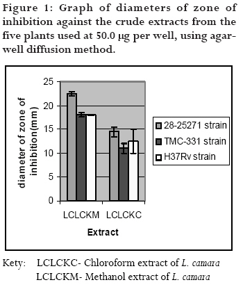
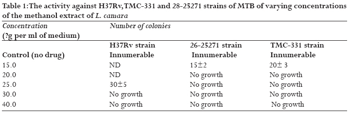
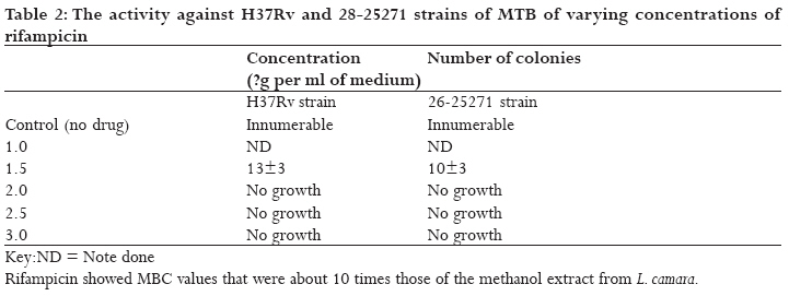
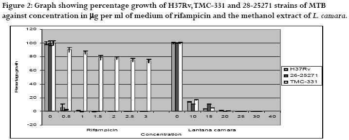
African Health Sciences, Vol. 9, No. 1, March, 2009, pp. 40-45 The anti-mycobacterial activity of Lantana camara a plant traditionally used to treat symptoms of tuberculosis in South-western Uganda 1Claude Kirimuhuzya, 1Paul Waako, 2Moses Joloba, 3Olwa Odyek 1Department of Pharmacology and Therapeutics, Makerere University, Code Number: hs09007 Abstract Introduction: Tuberculosis continues to be a devastating public health problem. Many communities in Uganda use medicinal
plants to treat various infections, including respiratory tract infections. There are claims that some can treat tuberculosis. Verifying some
of these claims could lead to discovery of lead compounds for development of a TB drug. Key words: Anti-mycobacterial; In vitro activity; Medicinal plant; Lantana camara; Mycobacterium tuberculosis; rifampicin Introduction Tuberculosis (TB) remains a devastating global public health problem in developing countries. Globally, more than one-third of the world's population (about 2 billion) is infected with the bacterium that causes TB. 1, 2, 3 Approximately 9 million people are diagnosed with the disease of which 2 million people die annually. 3, 4, 5 The emergence of multi-drug resistant strains of Mycobacterium tuberculosis and more recently extensively drug resistant tuberculosis (XDR TB) poses a formidable challenge to the control of the disease.2 Chemotherapy is the mainstay of tuberculosis control and there is need to develop new drugs for the control of tuberculosis, particularly the Multi-drug resistant (MDR) and XDR TB strains. There is also need to alleviate the shortcomings of current drug regimens by developing safer, more effective and more affordable agents that can act over shorter periods of time. Plants have been a source of effective chemotherapeutic agents for various infectious diseases and there is a growing interest in the development of drugs of plant origin. Many communities in East Africa, in general, and Uganda in particular, traditionally use plants to treat various infections, including respiratory tract infections. There are claims that some can, actually, treat tuberculosis. Lantana camara is one of the plants claimed to treat tuberculosis6 and is widely used in some parts of South-western Uganda. L. camara is a shrub that belongs to the family of Verbanaceae. It is reported to have its origins in the tropics of the Americas but is now found in many African countries including some arid regions, and is widespread in Kenya, Uganda and Tanzania after its introduction as ornamental plant about 300 years ago. 7 It is highly invasive and currently occupies a large percentage of the vegetation cover wherever it was introduced. Its growth poses a threat to other biodiversity. Various uses of L. camara have been reported. It is mainly used as herbal medicine but other uses like providing a source of firewood, mulch, making hedges, and use as a source of microbicides, fungicides and nematicides, insecticides have also been reported. 7 In traditional use in South-western Uganda, the leaves of the plant are chewed with rock salt and the extract swallowed, or they are crushed raw or baked, mixed with rock salt and the extract taken orally for the treatment of cough (Pers com). Chemical compounds isolated from extracts of leaves of L. camara are reported to have shown to exhibit antimicrobial, fungicidal, insecticidal and nematicidal activity. 7 There are also reports that lantana compounds isolated from the extracts can be applied as weed killers and have been tested on the water hyacinth with some success. 7 It is further reported that verbacoside, a compound isolated from lantana extract has been demonstrated to possess anti microbial immunosuppressive and anti tumour activities. 7 Use of lantana oil in treatment of skin itches and as an antiseptic for wounds and externally for leprosy and scabies is also reported. 7 Use of lantana extracts in folk medicine for the treatment of cancers, chicken pox, measles, asthma, ulcers, swellings, eczema, tumors, high blood pressure, bilious fevers, catarrhal infections, tetanus, rheumatism, malaria and atoxy of abdominal viscera, is also reported. 7 In this study we investigated the anti-mycobacterial activity of leaf extracts from L. camara. Materials and methods Plant collection and identification Leaves of L. camara were collected from Kagashe Village of Eastern Ward of Rukungiri Town Council in Rukungiri District, South-Western Uganda, approximately at Latitude 0.8oS and longitude 30oE. The team included a Taxonomist, Principal investigator and a Traditional Healer. Only plants judged as mature by the team were collected and the collection was done during the rainy season (March- May). Plants, which appeared to have viral, bacterial or fungal infections, were discarded. A shoot with leaves and flowers was used for identification and a voucher specimen (CKK005) was kept at the Makerere University herbarium. Extraction and preparation of test samples Leaves of L. camara were dried at room temperature away from direct sunshine. The dried leaves were pulverized, weighed and stored at room temperature. The powdered plant material (100 g) was percolated sequentially for four days with 500 ml of chloroform, methanol and water respectively. Filtration was done and the dry extract obtained by rotary evaporation at a temperature of 40oC under reduced pressure to a minimum volume and then drying at room temperature (8, 9). The water extract was got from the extracting solvent using a freeze drier. The extracts were then weighed and the yield determined. The dry extracts were stored at room temperature for further testing. Study strains of M. tuberculosis Strains of M. tuberculosis were obtained from the Joint Clinical Research Centre (JCRC), Mengo, Kampala, Uganda. Three strains were used in the study. They included the rifampicin-sensitive 28-25271, rifampicin- resistant TMC-331 strain and the H37Rv strain. H37Rv and TMC-331 are internationally used as standard MTB strains for pan sensitivity and rifampicin-resistance, respectively. The 28-25271 MTB strain was isolated from a Ugandan patient by JCRC. Culture and preparation of M. tuberculosis inoculum Stored strains were cultured using the Middle brook 7H11 (Difco®) agar supplemented with oleic acid-albumin-catalase (OADC). No adjustments for pH were made. The work was done in a high-class biological safety cabinet using the method of Pereira, et al (2005) with slight modifications (10). Confirmation of positive cultures was done using fluorescence microscopy with rhodamine-auramine stain. 11, 12 Antimycobacterial sensitivity testing The disc diffusion and agar dilution method was used. Dried methanol and chloroform crude extracts were dissolved in dimethylsulfoxide (DMSO) to a concentration of 50 mg/ml to make a stock solution. The stock solution was sterilized using 0.2 μm single use filters (Sterile Acrodic®). Sterile distilled water was used for further dilution. Water extracts were dissolved in sterile distilled water. A concentration of 50.0 μg per well was used for the general susceptibility tests. A stock solution of rifampicin, 3.0 mg/ml was prepared from a preserved frozen stock of a concentration of 20.0 mg/ml. A concentration of 1.0 mg/ml was used for susceptibility testing in the case of rifampicin. The medium for incubation was sterile Middle brook 7H11 agar in 90-mm diameter Petri dishes with quadrants. In each quadrant of the Petri dish was put 5.0 ml of the medium. The solidified medium in the quadrants was inoculated using the flooding method so that a uniform surface distribution of inoculum was obtained. Wells of 5.0 mm diameter and 2.5 mm depth were then bored in the dry inoculated medium using a sterile cork borer, and 50 μl of the test extract, were dispensed into the well of the first quadrant in each Petri dish giving an extract concentration of 50.0 μg per well. A volume of 50 μl of the 1.0 mg/ml solution of rifampicin and an equal volume of each of the plant extracts were dispensed into the well of the second quadrant giving a drug concentration of 1.0 μg per well. The well in the third quadrant was left empty as a control, while the well in the fourth quadrant was filled with the solvent used to dissolve the extract, also as a control. The Petri dishes were then left in the hood overnight to allow diffusion of the extracts and drug and then sealed with a carbon dioxide-permeable tape and incubated at 37°C in a carbon dioxide incubator for up to four weeks. Activity of extracts and rifampicin was determined from the zone of inhibition surrounding the well. The sensitivity of M. tuberculosis to the extracts and the drug was determined by measuring the zones of inhibition surrounding the well using a millimetre scale. The actual diameter of zone of inhibition was got after subtracting the diameter of the well. Three replicates of each test were done for each MTB strain. A rough Minimum Inhibitory Concentration (MIC) was determined for the methanol extract by dispensing serial dilutions of the test extract, concentrations ranging from 5.0 mg/ml to 50.0 mg/ml. McFarland No.1 standard was used in the preparation of the inoculum. Dilutions of rifampicin, using appropriate calculations, were also used for comparison. After incubation, the lowest concentration that inhibited growth of the bacteria on the medium, shown by absence of growth around the well, was taken to be an estimate of the MIC of the extract or the drug. Determination of Minimum Inhibitory Concentration (MIC) L. camara methanol extract concentrations were dispensed in the medium at intervals of 5.0 mg/ml to determine the actual MIC. The MIC was taken to be the concentration of the drug or extract that inhibited growth of M. tuberculosis by 99 percent or greater, in comparison to the positive growth control. Fresh cultures (up to 3 weeks old) were used as source of M. tuberculosis using a standardized inoculum prepared basing on McFarland No.1 standard, as in the case of the pilot study. The agar dilution method was used to get the definitive MIC value. Serial dilutions of the extracts and rifampicin were made and each solution added to the autoclaved medium cooled up to 50oC. The concentrations were made at 10 ml per ml of medium and 5 ml of the medium at each of the concentrations was then poured into quadrants corresponding to the concentrations in the Petri dishes. One quadrant in each Petri dish contained the medium with no drug as the growth control. The quadrants were then inoculated with 10-4 dilution of inoculum at McFarland standard No.1. Activity was then determined from the number of colonies in each quadrant. The concentration ranges used were spread around the estimated MIC and ranged from 10 mg/ml to 40 mg/ml. 13 After the period of incubation, the numbers of colonies on the drug containing quadrants were determined and expressed as percentages of those on the drug-free quadrant. Minimum Bactericidal Concentration (MBC) tests This was taken to be the concentration of the drug or extract that prevented growth by 99 percent or greater, compared with the untreated controls. To get the inocula for the MBC tests, the surface of the media in the drug-inhibited quadrants in the MIC test, were scraped and 10-fold dilution of inocula were prepared using the usual procedure of preparing inocula. Fresh Middle brook 7H11 in 90-mm plates with quadrants were then prepared, and inoculated with the inocula prepared above and the procedure for incubation repeated for up to eight weeks.Toxicity test on the crude methanol extract of L. camara in mice Acute toxicity test on the most active extract was done on mice, Mus musculus. It was carried out as described by Ghosh (1984) using mice of both sexes, for each concentration.14 The mice were fasted overnight and four dose ranges were used. Pairs of mice, using widely separated doses at 10, 50, 100 and 500mg/kg by oral route using a syringe fitted with a canula. Attempts were also made to study some acute drug effects for which it was possible to observe within twenty-four hours. Data collection and analysis The numerical data from the replicated investigations is presented in form of tables and histograms. Statistical analysis involved use of the statistics computer program, SigmaPlot, New Version 10, of Systat Software Inc. (2002). Ethical considerations The Research and Ethics Committee of the Faculty of Medicine, Makerere University and the Uganda National Council of Science and Technology approved the study. The animals used were handled in accordance with international guidelines for the handling of experimental animals. Results Estimation of antimycobacterial activity There was difficulty of diffusion of crude extracts as the distance from the well increased. Rifampicin showed no activity on TMC-331 at 1 μg per ml of medium but showed complete clearance of the quadrant for both 28-25271 and H37Rv strains. Water extracts hardly showed any activity at the concentrations used, and were not treated further. The chloroform and/or methanol extracts of L. camara, showed some activity against MTB strains used, and were treated further but at a higher concentrations ranging between 50 and 5 μg/ml. However, activity of rifampicin was more than 10-fold for the active extracts. For the solvents used, there was no sign of inhibitory activity on all the MTB strains used. Rifampicin completely cleared quadrants against H37Rv and the wild strain. The methanol extract of L. camara leaves showed the highest activity but it was much less active than rifampicin on H37Rv and the wild strain (28-25271. The chloroform extract (LCLCL) of L. camara also showed activity against all the three strains of MTB but was less active than its methanol (LCLCKM) counter part (fig. 1). The antimycobacterial activity of L. camara leaf extracts The methanol extract of L. camara showed an MIC value of 20 µg/ml for H37Rv and15 µg/ml of medium for TMC-331and the wild strain (28-25271), compared to rifampicin which showed MIC values of 1.0 µg/ml of medium for H37Rv and the wild strain, but was ineffective against the rifampicin-resistant TMC-331 strain of MTB. Rifampicin showed complete inhibition of growth for H37Rv and the wild strain at 1.5 µg/ml of medium but was unable to inhibit TMC-331 even at a concentration of 3.0 µg/ml of medium. This showed a clear contrast with the methanol extract of L. camara, which was active against all the strains of MTB used. The acute toxicity profile of the methanol extract of L. camara When pairs of mice (one male and one female) were given oral doses, in a pilot study using the methanol extract of L. camara, the median lethal dose was found to be greater than 500 mg/kg body weight. This was outside the range usually considered for acute toxicity. The mice experienced sedation for about six hours at a dose of 500-mg/kg-body weight but there were no anaesthetic or analgesic effects as the sedated animals could still respond to a pinch on the tail. There was also increased breathing rate and restlessness at doses of 100mg/kg body weight and above. However, all the animals showed normal activity after the twenty four hours of observations (tables 1, 2, figure 2). Discussion The methanol leaf extract of L. camara has the best activity among the three extracts investigated against three strains of M. tuberculosis; H37Rv, TMC-331and the wild strain, (28-25271). Rifampicin was found to be more than 10 times active than the methanol extract against the two of the study strains. The methanolic extract of L. camara showed activity against the rifampicin resistant strain. The leaf extract of this plant was found non-toxic to mice. L. camara is a common ornamental plant in many parts of East Africa. Studies have reported growing interest in its medicinal values. 7 The growth of this plant threatens the rest of the biodiversity in the region. 7 Previous studies have reported some compounds, which could be responsible for the biological activity. 7 We are reporting the antimycobacterial activity for the first time. With the existence of MDR, this is landmark discovery, more so when the extracts have been seen to be effective against the rifampicin resistant strains of M. tuberculosis. The comparatively low activity of the extracts of L. camara and the other extracts compared to rifampicin could be explained by the fact that the former are crude extracts while the latter is one of the most powerful among the anti TB drugs: pure compounds might have much higher activity. However, L. camara extracts showed an advantage over rifampicin by being highly active against the rifampicin-resistant strain of MTB. Since rifampicin resistance is a good indicator of MDR TB, these extracts could be active against MDR-TB. However, further studies need to be done to ascertain this. This study opens a possibility of obtaining novel compounds for the treatment of tuberculosis including MDR. We used in vitro systems in this study and earlier studies have reported variations in antimicrobial activity when the study is repeated in an in vivo system. There is, therefore, need to carry out some more studies in animal models of tuberculosis to reaffirm these findings. Furthermore, there could be need to investigate the part of the plant that shows better activity as well as carrying comprehensive phytochemical analysis of the plant extracts.. The documentation of the medicinal value of this notorious and rapidly growing plant brings a lot of relief to the plant conservation community, which is concerned about its effect on other biodiversity. In effect this plant has potential of being exploited for its medicinal value and hence controlling its ground cover in this region and the world at large. Acknowledgements We are grateful to the Uganda National Council for Science and Technology for granting permission to conduct this study. We also thank the staff and management of the Joint Clinical Research Centre, Mengo, Kampala for allowing us access to their laboratories. We feel greatly indebted to Dr. Kalema David of the Department of Botany, Makerere University for the taxonomic identification of the plant material References
Copyright © 2009 - Makerere Medical School, Uganda The following images related to this document are available:Photo images[hs09007f1.jpg] [hs09007t1.jpg] [hs09007f2.jpg] [hs09007t2.jpg] |
| |||||||||
