 |
| About Bioline | All Journals | Testimonials | Membership | News |
|
||||||
|
||||||
African Health Sciences, Vol. 10, No. 1, March, 2010, pp. 9-13 Calcium, inorganic phosphates, alkaline and acid phosphatase activities in breast cancer patients in Calabar, Nigeria *Nathaniel I. Usoro1, Maxwell C. Omabbe2, Chinyere A. O. Usoro2 & Augusta Nsonwu2 1Surgery Department University of Calabar Teaching Hospital, Calabar, Nigeria *Correspondence author: Dr. Nathaniel I. Usoro, Consultant General Surgeon, Surgery Department, University of Calabar Teaching Hospital, Calabar, Nigeria E-mail: natusoro@yahoo.com Phone: +234 8037028752 Code Number: hs10003 Abstract Background: Breast cancer is the commonest malignancy of women in Nigeria. Change in serum levels of
some biochemical parameters could assist diagnosis and follow-up of breast cancer.
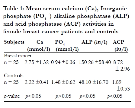
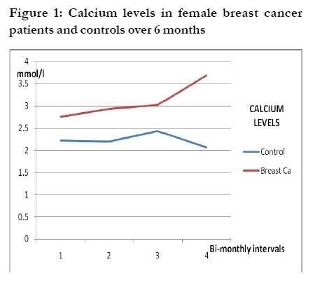
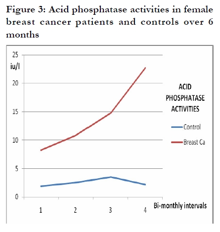
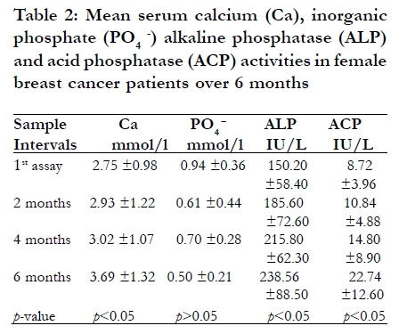
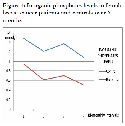
Key words: Breast cancer, serum calcium, inorganic phosphate, alkaline phosphatase, acid phosphatase, tumor markers. Introduction Breast cancer is the most frequently diagnosed malignancy and the second principal cause of death among women world wide as well as in Nigeria.1, 2, 3 The incidences of the disease appear to be on the increase in population groups that hitherto enjoyed low incidence.4 The increasing prevalence of breast cancer in our society has been attributed to changing life style and dietary pattern from the traditional African pattern to the western pattern. Breast cancer like any other disease condition is associated with derangement in the body's physiological functions, alterations in the homeostasis and production of some biochemical metabolites. Production of tumor markers has been utilized as a parameter for diagnosis and monitoring disease progress. Screening for markers such as CA 15-3, CA 549, CA 27-29 and mucin-like carcinoma-associated antigen (MCA) has been utilized for early disease diagnosis and intervention.5, 6 Some studies have shown increased serum levels of calcium and elevated activities of alkaline phosphatase (ALP) and acid phosphatase (ACP) in patients with malignancies including breast cancer.7, 8, 9, 10 The mechanisms postulated for the increased levels of these parameters in malignancy includes humoral mechanisms mainly mediated by parathyroid hormone related peptide (PTH-rp), osteolytic bone metastasis11 and increase in concentration of ACP particles which characterize lysosomes in breast tumor cells.7 Assessment of the serum levels of these biochemical parameters could substitute for the classical tumor markers in areas where facilities for these are not readily available. Calabar is one of such areas; hence this study is aimed at assessment of serum calcium, inorganic phosphates, ALP and ACP activities in breast cancer patients and change in the serum levels over time. Methods Patients and Study Design This case-controlled comparative study was carried out in the department of surgery and department of chemical pathology, University of Calabar Teaching Hospital, Nigeria. The patient's consent was sought before recruitment into the study. Twenty-five female breast cancer patients confirmed by biopsy were selected by convenience sampling. Twenty-five age-matched breast cancer-free females were used as controls after exclusion of the disease by history and clinical examination. None of the patients or control subjects was on calcium supplements. A follow up study was carried out on the women for six months with bi-monthly analysis of serum calcium, inorganic phosphates, ALP and ACP activities. Sample Collection Five milliliters of whole blood samples were collected from each subject into plain tubes with precautions to avoid pre-analytical errors, avoiding the use of tourniquet and fist clenching.12 Serum was extracted for analysis of total calcium and inorganic phosphate, and ALP and ACP activities. Total calcium and inorganic phosphate was estimated using colorimetric methods,13, 14 while ALP and ACP activities were determined using the enzymatic method.15, 16 Statistical Analysis Data was analyzed using the paired t-test analysis and analysis of variance (ANOVA) for inferential statistics between study group and control subjects. Results Serum calcium levels, ALP and ACP activities were significantly higher (p<0.05) in female breast cancer patients when compared with those of the control subjects. No significant difference (p>0.05) was seen in the inorganic phosphate levels of both groups as shown in Table 1. Serum calcium levels, ALP and ACP activities increased significantly in women with breast cancer with time (p<0.05) as shown in Figure 1, Figure 2, and Figure 3, and Table 2, whereas no significant variation (p>0.05) was observed in the inorganic phosphate levels with time (Figure 4 and Table 2). There was no significant variation (p>0.05) in the levels of serum calcium, inorganic phosphate, ALP and ACP activities in the control group during the 6 months follow up (Figure 1, Figure 2, and Figure 3 and Figure 4). Discussion The results of this study have shown that women with breast cancer have higher total serum calcium levels and higher ALP and ACP activities when compared with the control subjects. Hypercalcemia and high ALP and ACP activities have also been reported in other malignancies.9, 17, 18, 19, 7, 10 The hypercalcemia in breast cancer has been attributed in part to osteolytic bone metastases and this account for 20-30% of the hypercalcemia cases in oncology patients. The skeletal invasion and destruction by tumor induced by tumor-production of various cytokines such as transforming growth factor-α (TGF-α), tumor necrosis factor-α (TNF-α), TNF-β, interleukin-1 and interleukin2, leads to increasing bone osteolysis20 and modification of the reabsorption, excretion and resorption of calcium and phosphate ion.21 Alteration in humoral regulation of calcium resulting from production of parathyroid hormone related protein (PTH-rp) has also been implicated in tumor associated hypercalcemia.22, 23 Plasma concentration of PTH-rp is rarely elevated in healthy individuals, but elevated concentrations are detectable in about 80% of hypercalcemia patients with solid tumors. PTH-rp interacts with parathyroid hormone receptors on cell membranes, activating adenyl cyclase, which triggers an increase in cyclic AMP production and increases intracellular calcium. These actions are responsible for increasing bone demineralization and elevating serum calcium concentrations, decreasing reabsorption of phosphate in the proximal renal tubules, increasing calcium reabsorption in the distal tubules and increasing cholecalciferol production.17ACP activity has been used to indicate that lysosomes participate in the execution of cell death in a variety of tissues and the regression of mammary carcinomas.7 Activities of lysosomal acid hydrolases have been demonstrated to be more marked in cancer cells than in homologous normal tissue24, 25 and the histochemical pattern of ACP distribution in the breast tissue showed differences between normal and neoplastic cells.7 Significantly elevated tartrate resistant ACP 5b activity was also reported by Chao & co-workers, (2005).26 ACP has been used as a marker of metastatic bone disease and response to treatment in breast cancer patients.27 ALP has many isoenzymes localized in the liver, bones and in lesser amounts the intestines, placenta kidney and leucocytes.28 Another isoenzyme of ALP termed Regan isoenzyme has also been identified in various malignancies29 and this may be contributing to increased ALP activity seen in breast cancer patients. The increased activity of this enzyme seen in subjects of the study may also be due to osteolytic bone metastases in breast cancer leading to increased osteoclastic activity and bone resorption. Increase in serum ALP levels is however non-specific as it is also frequently associated with a variety of other diseases. Also, the elevation of ALP activity to less than three times the normal level is usually not considered significant.9, 30 Conclusion Women with breast cancer have higher calcium levels and higher ALP and ACP activities than normal healthy women. The progressive increase in the serum calcium levels and ALP and ACP activities during the six months follow up study is an indication that measurement of these parameters may be useful tools in monitoring treatment and disease progression in areas where facilities for sophisticated studies are not readily available. Acknowledgement The assistance of Mr Henry Efobi of Chemical Pathology Department, University of Calabar Teaching Hospital, Nigeria, in respect of a citation is appreciated. References
The following images related to this document are available:Photo images[hs10003t1.jpg] [hs10003f3.jpg] [hs10003t3.jpg] [hs10003t2.jpg] [hs10003f4.jpg] [hs10003f2.jpg] [hs10003f1.jpg] |
| |||||||||
