 |
| About Bioline | All Journals | Testimonials | Membership | News |
|
||||||
|
||||||
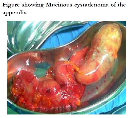
African Health Sciences, Vol. 10, No. 1, March, 2010, pp. 99-100 Case report Mucinous cystadenoma of the appendix: a case report *Alese OB, Irabor DO Division of Gastrointestinal Surgery, Department of Surgery, University College Hospital, Ibadan, Nigeria *Corresponding author: Olatunji B. Alese, Department of Surgery, University College Hospital, PMB 5116, Ibadan, Nigeria Tel: +234 803 4058250, 2410088 Ext. 2500 E-mail: aleseob@yahoo.com Code Number: hs10018 Introduction Tumours of the appendix are emerging as diseases of increasing concern due to a rising incidence1. We present a case of mucinous cystadenoma of the appendix in an elderly patient. To our knowledge, this is the first report of mucinous cystadenoma of the appendix from Nigeria. Key Words: Appendiceal tumour, Appendicectomy. Case report: A 74 year old man presented with a one-month history of recurrent episodic dizziness and occasional excessive sweating. The dizziness was precipitated by turning his neck to the right side. He presented to the hospital after the third episode of fainting spells where he was seen at the medical out-patients clinic. Examination revealed an elderly man who was afebrile, and was not pale. His blood pressure was 145/90mmhg; pulse rate was 78/minute, regular, with some hardening of his radial artery. Both lung fields were clear on auscultation His abdomen was flat and soft with a firm sausage-shaped non-tender and non-mobile mass in the right iliac fossa. The clinical diagnosis was caecal tumour; differential diagnosis of carcinoid syndrome. Abdominal ultrasonography revealed a 21cm x 6cm x 7.5cm septate, cystic mass in the right pelvis, and a normal liver architecture. ECG revealed supraventricular extrasystoles. Cervical X-rays revealed prominent osteophytes on the cervical vertebrae. He was subsequently prepared for an exploratory laparotomy. Intra-operative findings included scanty serous ascites, a sausage shaped, firm appendiceal tumour, which was not attached to the surrounding structures as shown in the Figure below. He had a right hemicolectomy. The tumour weighed 480g. The histology of the appendix was reported as `showing extensive pools of mucin in a cystic cavity lined by tall columnar epithelium with apical mucin. Features are those of mucinous cystadenoma of the appendix'. His postoperative recovery was delayed due to prolonged ileus. He however resumed bowel function by the 9th postoperative day. His clinical status thereafter was unremarkable, and he was discharged home on the 13th postoperative day. Discussion Mucinous cystadenoma is a rare tumour of the appendix associated with cystic dilatation, to which the more general term of mucocele has been applied2. Mucocele of the appendix denotes an obstructive dilatation of the appendiceal lumen due to abnormal accumulation of mucus, which may be caused by a retention cyst, mucosal hyperplasia, cystadenoma and cystadenocarcinoma3, 4. It is a rare entity found in 0.3% of appendiceal specimens5, 6, with a slight female predominance and an average age at diagnosis of over 50 years7. Mucocelesare often asymptomatic and are discovered as incidental findings at appendicectomy6, during laparotomy for another indication, or during histologic examination of an operative specimen8. However, it may be diagnosed clinically from features of acute appendicitis8. Association with concomitant colon cancer is recognized9. A link with irritable bowel disease (IBD) has been reported and IBD is a known risk factor for colorectal neoplasia10. It may also be an incidental finding during ultrasonography, computed tomography, and other radiological examinations of the gastrointestinal tract. Four pathological entities are described from the epithelial characteristics: (1) Simple or retention mucoceles due to obstruction of the appendiceal outflow; usually by a faecolith, characterized by normal epithelium and mild luminal dilatation (2) Mucoceles with hyperplastic epithelium with mild luminal dilatation. These constitute 5%-25% of mucoceles. (3) Mucinous adenoma/cystadenoma is the most common form, accounting for 63%-84% of cases. These exhibit mostly epithelial villous adenomatous changes with some degree of epithelial atypia. There is marked distention of the lumen up to 6 cm. (4) Malignant mucinous cystadenocarcinomas, represent 11%-20% of cases. These demonstrate glandular stromal invasion and/or presence of epithelial cells in the peritoneal implants.11 There is always the risk of rupture, either spontaneous or accidental, with consequent development of pseudomyxoma peritonei2,11, 12, which may present with features of intestinal obstruction13. Treatment is usually an appendicectomy; right hemicolectomy is performed when the caecum is involved8,11,12, 14, 15. Our patient has been seen for follow-up thrice at the surgical out-patients clinic; initially at 2 weeks post-discharge, then 6 months and then after 1 year. At the time of this report he was in good health. References
Copyright © 2010 - Makerere Medical School, Uganda The following images related to this document are available:Photo images[hs10018f1.jpg] |
| |||||||||
