 |
| About Bioline | All Journals | Testimonials | Membership | News |
|
||||||
|
||||||
African Health Sciences, Vol. 12, No. 1, March, 2012, pp. 63-68 Facial approximation: evaluation of dental and facial proportions with height * Esan TA1, Oziegbe OE2, Onapokya HO3
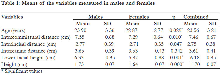
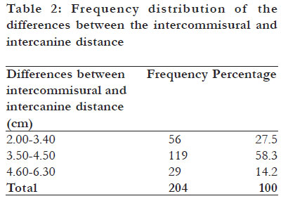
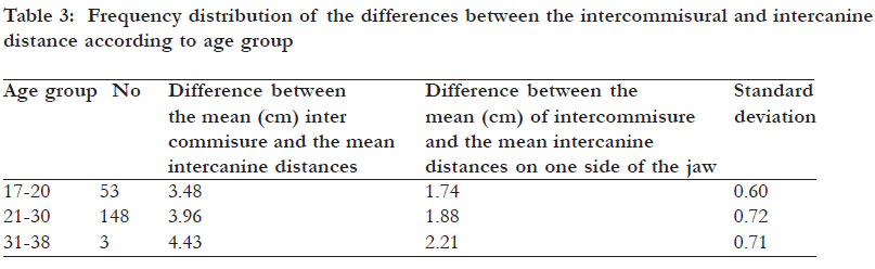
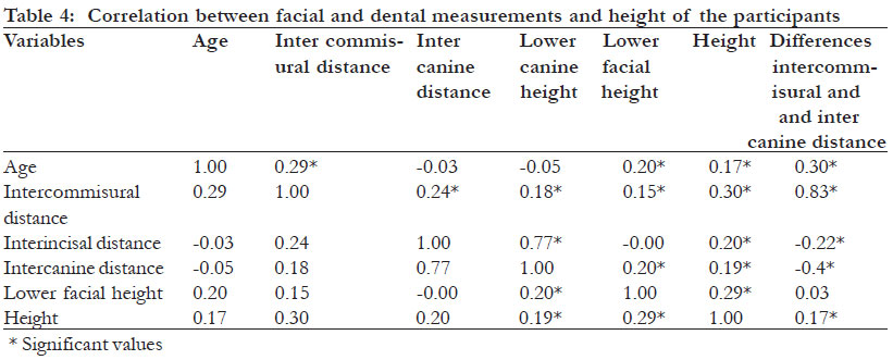
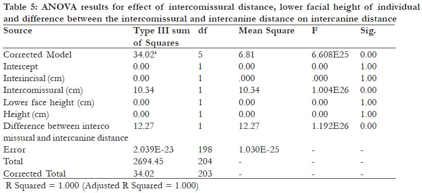
Code Number: hs12011 Abstract Background: Fabrication of complete dentures requires the use of certain guidelines which are placed on the bite blocks to assist the clinician to have the maxillary anterior teeth restored to optimal dento-labial relations, in harmony with the overall facial appearance. Key words: facial measurements, intercanine , intercommisure Introduction Esthetics is of primary concern for patients seeking complete removable prosthesis. The goal of treatment is to have the maxillary anterior teeth restored to optimal dento-labial relations in harmony with the overall facial appearance. In order to achieve this, the dimension, morphology and arrangement of the anterior teeth must be in proportion to the facial dimension. During bite registration in complete denture fabrication some guidelines such as smile line, center line and canine line are placed on the bite blocks in order to assist the clinician in determining the correct size of the anterior teeth. These lines are placed based on reference to certain facial land marks such as intercommissure, interalar, the labial frenum, bizygomatic width and interpupillary distance.1-5 Certain authors have proposed a relationship between the width of the maxillary central incisor and the interpupillary distance.2,4 Cesario and Latta2 found that a ratio of 6.6 which had previously been proposed existed between the interpupillary distance and the central incisor width in white men and women, and also in black women. Similarly, a proportional relationship between the widest part of the nose and the anterior dental arch has been reported.3,6 The relationship between the width of a central incisor and the bizygomatic width (1:16) is commonly used to determine the size of the maxillary anterior teeth.7-9 Recently in a study by Latta et al4 the relationship among the width of the mouth, the inter alar width, the bizygomatic width, and the interpupillary distance were evaluated. It was concluded that these relationships might be used as references if applied in combination, although racial and gender differences were detected when anatomic measurements were evaluated individually. In Nigeria, there are no studies linking facial proportions with the size of anterior maxillary teeth that could be used as guide for determining/ defining the maxillary anterior dentolabial dimensions. The usual practice has been the reliance on some values and guidelines established among Caucasians and Asians. Therefore, this study aimed to explore if any relationship exists between facial proportion and height of individuals with their dental proportions. Methods This was a crosssectional study involving 204 dental students of Obafemi Awolowo University, Ile-Ife, Nigeria who volunteered to participate in the study. The students were from south-western Nigeria. Written informed consent was obtained from them. The study was explained to all the participating dental students and they were asked to report to one of the authors (EOO) to ascertain suitability for inclusion and subsequent enumeration. Consenting students with previous facial trauma/ surgery, tooth loss in the anterior segment, those wearing dentures, crowns/bridges in the anterior segment, and those with history of neurological diseases were excluded. Demographic information such as the age and gender of each student were obtained. The height of each student was measured using a standiometer as the maximum distance from the floor to the highest point on the head with the student facing directly and looking ahead. The shoes were off with the feet together, arms by the sides, heels, buttocks and upper back in contact with the wall. Lower facial height was measured by asking the participants to be seated on a dental chair with their Frankfort plane parallel to the floor. With the aid of a Willis gauge the distance between the septum of the nose and the chin of each participant was measured as the lower facial height. The interincisal and intercanine distances were measured by asking the student to bite into a sheet of tough modeling wax. The inter incisal distance was measured as the distance between the disto proximal surface of the indentation of maxillary right permanent lateral incisor and the disto proximal surface on the indentation of maxillary left permanent lateral incisor on the tough modeling wax. The intercanine distance was also measured as the distance between the distoproximal surface on the indentation of maxillary right permanent canine and the distoproximal surface of the indentation of the maxillary left permanent canine on the tough modeling wax. Where the indentation was not visible enough the student was asked to repeat the process. Three measurements were made for each variable studied by one of the authors and the average values of lower facial height, inter canine distance and inter incisal distance were calculated for each student. Also with the student relaxed and with the teeth in occlusion, the intercomissural distance was measured by a flexible meter rule. The measurement was performed three times by the same author and the average value was calculated. The data were imputed, analyzed and reported as simple frequency, means and standard deviations using the SPSS vs 11. Gender based comparison of parameters was conducted using the student t-test; correlation coefficient were determined between height and the parameters and statistical significance inferred at p<0.05. Correlation coefficient r <0.3 was considered as weak, 0.3-0.5 as moderate, >0.5 – 0.7 as significant correlation, >0.7-0.9 as high correlation, and >0.9 as very high correlation. Results The age range of the 204 students was from 17 - 38 years with the mean age being 23.56 + 3.21 years. There were 136 (66.67%) males and 68 (33.33%) females. The mean age for males was 23.90+ 3.36 years, while it was 22.87 +2.77 years for females. A statistically significant difference was found in the mean ages of males and females. (p=0.03) as shown in table I. The mean height of the total population was 1.70 + 0.07m. The males had significantly higher height (1.73 + 0.07m) than the females (1.64 + 0.07m) (p=0.000). The mean height of the males was significantly higher than the females when compared age for age. The mean intercomissural distance for the total population was 7.46 + 0.67cm. The mean intercomissural distance for male (7.55 + 0.68cm) was significantly more than the females (7.29 + 0.64cm, p= 0.01) as shown in table I. When the subjects were matched for age, the mean intercomissural distance for the males was higher than the females. The means of the interincisal and intercanine distance were 2.75 ± 0.38cm and 3.61 ± 0.41cm respectively for the total population. However, there was no significant difference between males and females. The mean of the lower facial height was 6.18 + 0.95cm for the total population. It was 6.33 + 0.95cm for males, while it was 5.87 + 0.88cm for females. The difference in the means was statistically significant with the males having higher values than females. (p<0.001) as shown in table 1. Table 2 showed that for a majority of the respondents (58.3%) the differences between the intercomissural and intercanine distance was between 3.50-4.50cm with the mean value, being 3.85± 0.72cm. The mean difference between the intercomissural and intercanine distance was 3.48cm for the age group 17-20years, 3.96cm for the age group 21-30 years, and 4.43cm for the age group 31-38years as indicated in table 3. There was a significant correlation between intercanine and interincisal distances as well as between the intercomissural distance and the differences between the intercomissural and the intercanine distances(r=0.8) while there were weak correlation between the height and intercomissural distance, height and intercanine distance, intercomissural distance and age (r=0.3 respectively). The correlation between lower facial height and age, interincisal distance, intercomissural distance and interincisal distance and height were low (r= 0.2 respectively) as indicated in table 4. Only intercomissural distance and the differences between intercomissural distance and intercanine distance had a significant relationship with the intercanine distance, as shown in table 5. Discussion A perfect smile is an important component of esthetics and this goes beyond having white and straight teeth. The smile should also be in proportion with the rest of the face. The proportion of facial structures and the relationship between facial measurements and natural teeth could be used as a guide to achieve this. One of the most difficult aspects during the selection of maxillary anterior teeth for a removable complete prosthesis is determining the appropriate mesiodistal width of the six maxillary anterior teeth.10 Many attempts have been made to establish methods of estimating the combined width of these anterior teeth, and improving the esthetic outcome. In earlier studies,8,11 measurements were made using extracted teeth. However, recent studies1215 attempted to measure the clinical tooth dimensions either on casts or using computer-based images or intraoral evaluations. When the values of the tooth widths as measured on the casts and anterior tooth images were compared, marked differences were found between the actual and perceived dimensions of the anterior teeth. This discrepancy was created by the curvature of the arch and angulations of the maxillary anterior teeth, in relation to the frontal plane of the photograph. Hence, tough modeling wax was used to make impressions of the anterior maxillary segment and then measurement of the impression of the distal point of one tooth to another was done with a caliper Nigeria is a diverse country with three major ethnic groups, the Yoruba, Hausa, and Ibo with each having different facial profiles. There is no dentofacial norm for use in prosthodontics that is representative of the Nigerian population. The present study aimed to determine if proportional relationships exists between the widths of the maxillary incisors, the intercanine distance, intercomissural distance, lower facial height, and the height of the individual in south western Nigerians. The study showed that males have a significantly higher mean value in all the parameters measured than the females. This is similar to other studies8,12,14 in which gender variations were noted in the dimensions of the anterior teeth for most racial groups, with men exhibiting wider anterior teeth than women. The reason may be due to the bigger physique of the males compared to the females in this study irrespective of the age group. In this study, none of the means of the various parameters measured was coincident. The mean intercomissural distance was greater than the mean of the intercanine distance measured. This is similar to the findings of Stephan,16 in which the mouth width differed significantly from the intercanine width. It was noted that the differences between these distances increased with age, with a majority of the participants having a mean difference of 3.50-4.50cm (1.75-2.25cm on one side of the jaw). Thus, these distances maybe a measure of growth/age of the individual. In the past, the distal surface of the natural upper canine was considered to be located near the corner of the mouth and this has been equated to the intercanine distance.17 This has been used in the selection of maxillary anterior teeth during bite registration. However, there are no evidences to suggest the validity of this relationship. Various authors2-5 have tried to find a relationship between the maxillary anterior teeth and facial structures. The interpuplillary line has been considered to be related to the intercanine line and has been widely used among Caucasians for the selection of maxillary anterior teeth. Since there are variations in facial proportions based on racial differences there is the need to investigate if a relationship exist between the intercanine distance and the interpupillary distance for other racial groups. The interincisal distance correlates strongly with the intercanine distance (r=0.774). Thus, the intercanine distance can be use to predict the interincisal distance. However, it is logical to expect this since the intercanine distance is one tooth away from the interincisal distance. Additionally, the study did not find any direct relationship between the intercanine distance and other parameters measured, except the interincisal distance. However, univariate analysis of variance showed a significant relationship between the intercanine distance and intercomissural distance on one hand, and the difference of the intercomissural and intercanine distances. This relationship should be explored in future studies. The mean lower facial height was markedly different from the intercanine and intercomissural distances. This may strongly indicate that these measurements can not accurately predict the width of the maxillary anterior teeth. Consequently, these guidelines should not be used in facial approximation to predict intercanine distance. It could not be determined in this study if the mouth width was equal to the distance between the mandibular second premolar. It was observed that inter commissural distance in a majority of the participants was about 1.75-2.45 cm distal to the intercanine distance on each side. This point is approximately close to the distal aspect of the second premolar. The relationship between the intercomissural distance and the second premolars seem probable since these teeth appear to be positioned closer to the cheilion than the canines.16 Conclusion The study determined the mean measurements of some facial parameters among Nigerians. This adds to the growing body of evidence that previously untested yet commonly used methods of facial approximations are considerably inaccurate. The study showed no direct relationship between intercomissural distance, interincisal distance, lower facial height, and the height of the participants with the intercanine distance. Therefore, intercomissural distance should not be used in marking canine line during bite registration procedure. At best, 1.75-2.45 cm should be subtracted from the intercomissural distance in order to determine the intercanine distance. References
Copyright 2012 - African Health Sciences The following images related to this document are available:Photo images[hs12011t1.jpg] [hs12011t5.jpg] [hs12011t4.jpg] [hs12011t2.jpg] [hs12011t3.jpg] |
| |||||||||
