 |
| About Bioline | All Journals | Testimonials | Membership | News |
|
||||||
|
||||||
Indian Journal of Surgery, Vol. 65, No. 3, May-June, 2003, pp. 277-279 Case Report Duodenal tuberculosis presenting as gastric outlet obstruction S. K. Raul, N. A. Das, R. Vakil, Y. D. Paljor, S. C. Joseph Department of Surgery, St. Stephen's Hospital, Delhi, India.
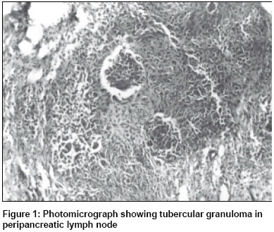
Paper Received: November 2001. Paper Accepted: March 2002. Source of Support: Nil Code Number: is03055 ABSTRACT Three cases of duodenal tuberculosis are presented. All had features of gastric outlet obstruction. Histopathological diagnosis of tuberculosis was obtained in two cases following endoscopic biopsy. Duodenal tuberculosis should be considered in patients presenting with gastric outlet obstruction, especially in areas where tuberculosis is endemic. How to cite this article: Raul SK, Das NA, Vakil R, Paljor YD, Joseph SC. Duodenal tuberculosis presenting as gastric outlet obstruction. Indian J Surg 2003;65:277-9. INTRODUCTION Gastrointestinal tuberculosis is an important health problem in developing countries. However, with the advent of the acquired immune deficiency syndrome, its incidence in the developed countries is also on the rise.1 Ileo-caecal and ileal are the usual forms seen in gastrointestinal tuberculosis. In the recent years, more and more cases of gastroduodenal tuberculosis are being reported. In areas where tuberculosis is endemic, diagnosis of duodenal tuberculosis must be kept in mind, particularly in patients with upper gastrointestinal obstruction and in those with peptic ulcer-like symptoms not responding to medical therapy. Case 1 A 25-year-old man in poor general condition presented with features of gastric outlet obstruction of 6 months duration. Upper gastrointestinal endoscopy showed a dilated stomach with food residue and an ulcer at the junction of the 1st and 2nd part of the duodenum. Ultrasound examination of the abdomen showed large mesenteric nodes. Barium studies revealed a narrowing between the 1st and 2nd part of the duodenum. Biopsy of the duodenal ulcer showed tubercular enteritis. A gastrojejunostomy with vagotomy was performed. Follow-up after 6 months on antitubercular treatment revealed no evidence of stricture in the 1st part of the duodenum. Case 2 A 31-year-old man was admitted with symptoms of gastric outlet obstruction of 2 months duration. Gastroduodenoscopy showed prepyloric swelling with an ulcer over the 1st part of the duodenum. Ultrasound of the abdomen revealed multiple mesenteric lymph nodes. Laparotomy revealed a dilated stomach with a stricture in the 1st part of the duodenum. There were multiple soft caseating nodes in the mesenteric, peripancreatic and para aortic regions. Gastrojejunostomy with truncal vagotomy was performed. Biopsy of the mesenteric node was reported as tubercular lymphadenitis. Follow-up endoscopy after 6 months on antitubercular therapy did not reveal stricture in the 1st part of the duodenum. Case 3 A 23-year-old woman presented with features of gastric outlet obstruction of 2 months duration. Upper gastrointestinal endoscopy showed a stricture at the junction of the 1st and 2nd part of the duodenum. Ultrasound of the abdomen showed numerous peripancreatic nodes. Endoscopic biopsy showed tubercular enteritis. Surgery was undertaken at which a duodenojejunostomy performed and a suprapancreatic lymph node was biopsied. Histopathology of the node showed tuberculous lymphadenitis. Follow-up endoscopy at 6 months revealed normal duodenum with no evidence of stricture. DISCUSSION Gastrointestinal tuberculosis is still rampant in developing countries and can mimic other gastrointestinal disorders. Isolated duodenal tuberculosis, without involvement of the other parts of the gastrointestinal tract does occur, though infrequently.1 The presentation of duodenal tuberculosis is varied; the commonest being gastric outlet obstruction.2-4 It may present as an acute emergency with bleeding or duodenal perforation. Obstructive jaundice may follow duodenal tuberculosis. The total number of cases reported till date amount to only about 50 (Ovid Medline till 2000). Most of the literature on duodenal tuberculosis is from tropical countries, especially India and South East Asia. There have been only occasional case reports from western countries like Italy,4 Spain,5 Germany6 and Canada.7 Proximal duodenal obstruction due to tuberculosis can masquerade as duodenal ulcer.2 Although the commonest cause of duodenal obstruction remains post-ulcer stenosis, atypical presentation of primary aorto-duodenal fistula caused by duodenal tuberculosis without an abdominal aortic aneurysm has been reported as an occasional case of pyloroduodenal fistula.8 Treatment in duodenal tuberculosis has been directed at the specific complication at presentation followed by appropriate antitubercular therapy.9 All the three cases reported by us presented with features of gastric outlet obstruction. None of the cases had any evidence of pulmonary tuberculosis. Truncal vagotomy with gastrojejunostomy was performed in two cases as there was insufficient length of dilated duodenum for anastomosis and acid peptic disease could not be ruled out. Duodenojejunostomy was performed in the other case. Duodenal tuberculosis is indeed a diagnostic challenge. Clinical evaluation, radiology and endoscopy10 are important modalities for diagnosis but they do have limitations. Despite the above modalities, sometimes it is extremely difficult to make a diagnosis and surgical intervention is required. Surgery is primarily directed at the presenting complication followed by a full course of antitubercular therapy. The importance of duodenal tuberculosis as a possible diagnosis in cases of gastric outlet obstruction in endemic areas of tuberculosis is hereby stressed. REFERENCES
Copyright 2003 - Indian Journal of Surgery. Also available online at http://www.indianjsurg.com The following images related to this document are available:Photo images[is03055f1.jpg] |
| |||||||||
