 |
| About Bioline | All Journals | Testimonials | Membership | News |
|
||||||
|
||||||
Indian Journal of Surgery, Vol. 65, No. 4, July-Aug, 2003, pp. 377-378 Case Report An unusual case of metaplastic breast carcinoma (sarcomatoid variant) Kiran Alam, Veena Maheshwari, Hasan Harris,* Ghazala Mehdi Departments of Pathology & *Surgery, Jawaharlal Nehru Medical College, Aligarh Muslim University, Aligarh 202002, Uttar Pradesh.
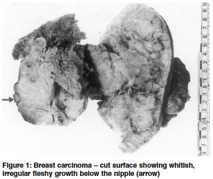
How to cite this article: Alam K, Maheshwari V, Harris H, Mehdi G. An unusual case of metaplastic breast carcinoma (sarcomatoid variant). Indian J Surg 2003;65:377-8. Paper Received: June 2002. Paper Accepted: April 2003. Source of Support: Nil Code Number: is03078 Abstract We present a case of a female aged 40 years with metaplastic carcinoma of the breast, which is a rare neoplasm. Although it is a tumour of the ductal type the predominant component of the neoplasm has an appearance other than epithelial or glandular and more in keeping with other cell types. Metaplastic sarcomatoid carcinoma runs a very aggressive course metastasizing more frequently via the blood stream than the lymphatics and bears an unfavourable prognosis for the patient. Key words: Metaplastic carcinoma, Neoplasm, Breast. Introduction Metaplastic carcinoma of the female breast is an uncommon lesion that may metastasize to body parts.1 There is confusion regarding the classification and staging of this tumour because of small number of cases seen. Its histogenesis is unknown.2 It may manifest as well-circumscribed or irregular spiculated masses. We present a patient with a lump in the breast diagnosed to have a metaplastic carcinoma of the breast. Case report A 40-year-old lady presented with a left-sided breast lump for 10 months. The lump had increased in size and had ulcerated over the preceeding 3 months. On examination there was a 10 x 6 cm, fungating-ulcerated growth in the left breast involving the nipple. It was hard in consistency and fixed to the breast tissue and the skin. The underlying muscles and the chest wall were free from the growth. The axilla had two enlarged but mobile lymph nodes. The general and systemic examination was normal except that she had moderate pallor. Apart from a haemoglobin of 8.5 gm%, the hematological and biochemical parameters were normal. X-ray of the chest and abdominal ultrasonography were normal. A fine needle aspiration cytology (FNAC) and imprint cytology showed sheets of anaplastic cells. The patient underwent a simple mastectomy with axillary sampling and postoperatively received chemo-radiation therapy. The resected specimen measured 16 x 12 x 6 cm and the ulcerated growth was 12 x 8 cm. The cut surface was whitish and irregular and there was a fleshy growth having a wide area below the nipple (Figure 1). The ulcer had necrotic debris. Microscopically, the tumour showed large areas of inflammation and necrosis. It resembled a sarcomatoid carcinoma, a variant of metaplastic carcinoma of the breast having spindle sarcoma-like stroma with osseous metaplasia. At places only a transition from ductal carcinoma to sarcoma-like element was seen (Figure 2). Blood vessel invasion was also present. Based on these findings, a diagnosis of metaplastic carcinoma of the breast with sarcomatoid variant was made. Discussion Metaplastic carcinoma of the breast is a rare entity, accounting for only about 0.02% of all breast carcinomas.2 The diagnosis can be difficult to establish both on clinical and histopathological bases and the behaviour seems to be more aggressive than that of the ordinary invasive ductal carcinoma. Broadly, there are four variants of metaplastic breast carcinoma: the sarcomatoid type, the spindle cell type, carcinoma in osteoclastic giant cells and squamous cell carcinoma. In the sarcomatoid type of metaplastic breast carcinoma, which was the variety encountered in our patient, there is a gradual transition from carcinomatous to sarcoma-like elements. The median age of presentation of metaplastic carcinoma of the breast in a series of 8 patients was 52.5 years3 and it was 50.5 years in another series of 14 cases.4 Our patient was significantly younger. Most of these tumours described were well-circumscribed lesions but our patient presented with an ulcerated mass which is rare, highlighting the late reporting of patients with breast malignancy in developing countries. The prognosis is poorer in this variety of metaplastic carcinoma than in the other types and the metastasis tends to be haematogenous rather than the more common route of the lymph node spread. Also, the best predictor for survival is the size of the neoplasm at the time of initial excision.5 It is therefore recommended that the malignant breast aspirates be carefully scrutinized for multiple breast components as metaplastic breast carcinoma, especially the sarcomatoid variety, can have a very aggressive course. References 1. Selvaggi SM, Kissner D, Qureshi F. Metastatic metaplastic carcinoma of the breast: diagnosis by bronchial brush biopsy. Diagn Cytopathol 1989;5:396-9. 2. Smith DM, Rongaus VA, Wehmann TW, Agarwal PJ, Classen GJ. Metaplastic breast carcinoma. J Am Osteopath Assoc 1999;96:419-21. 3. Kuo SH, Chen CL, Huang CS, Cheng AL. Metaplastic carcinoma of the breast: analysis of eight asian patients with special emphasis on two unusual cases presenting with inflammatory type breast cancer. Anticancer Res 2000;20:2219-22. 4. Chao TC, Wang CS, Chen SC, Chen MF. Metaplastic carcinomas of the breast. J Surg Oncol 1999;71:220-5. 5. Kaufman MW, Marti JR, Gallager HS, Hoehn JL. Carcinoma of the breast with pseudosarcomatous metaplasia. Cancer 1984;53:1908-17. © 2003 Indian Journal of Surgery. Also available online at http://www.indianjsurg.com
The following images related to this document are available:Photo images[is03078f2.jpg] [is03078f1.jpg] |
| |||||||||
