 |
| About Bioline | All Journals | Testimonials | Membership | News |
|
||||||
|
||||||
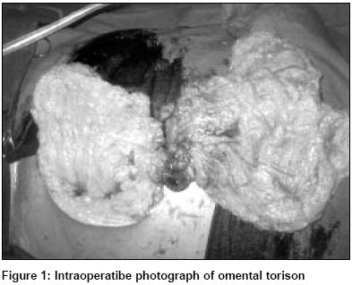
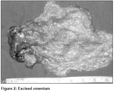
Indian Journal of Surgery, Vol. 65, No. 5, Sept-Oct, 2003, pp. 448 Images Omental torsion C. Rajesh Ballal, Rajeev P. Premnath Department of General Surgery, Kasturba Medical College, Mangalore.
Paper Received: March 2003. Paper Accepted: March 2003. Source of Support: Nil. How to cite this article: Ballal CR, Premnath RP. Omental torsion. Indian J Surg 2003;65:448. Code Number: is03096 A 35-year-old man presented with pain in the left hypochondrium of 4 days duration. There were no relieving or aggravating factors and no relation to food. He had no history of vomiting, bowel or bladder disturbances. He gave a past history of failed orchidopexy on the right side several years ago. On examination he had tenderness in the left hypochondrium and bilateral undescended testes. He had normal blood investigations and ultrasound abdomen showed a left inguinal hernial sac with omentum. CT scan of the abdomen showed the left testis at the left deep inguinal ring and thickened omentum. He underwent an exploratory laparotomy for persistent pain. We found an omental torsion on laparotomy (Figure 1), which was excised and sent for histopathology (Figure 2). No other pathology was found per abdomen. It was decided that left-sided orchidopexy be done at a later date. The patient had an uneventful postoperative recovery. Histopathology of the excised omentum was unequivocal. © 2003 Indian Journal of Surgery. Also available online at http://www.indianjsurg.com The following images related to this document are available:Photo images[is03096f1.jpg] [is03096f2.jpg] |
| |||||||||
