 |
| About Bioline | All Journals | Testimonials | Membership | News |
|
||||||
|
||||||
Indian Journal of Surgery, Vol. 66, No. 2, Mar-Apr, 2004, pp. 105-106 Case Report Keratinising desquamative squamous metaplasia of the upper urinary tract: A case report Sujata Nayak, Shaila C. Puranik, Vasudev V. Holla Department of Pathology, B. J. Medical College and Sassoon General Hospitals,
Pune - 411001, India.
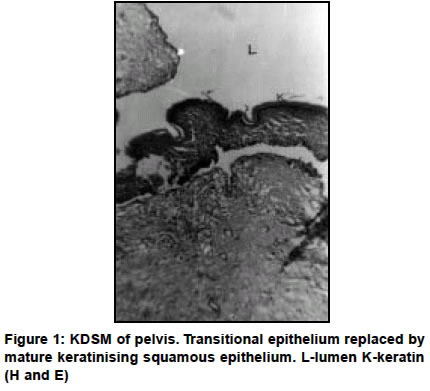
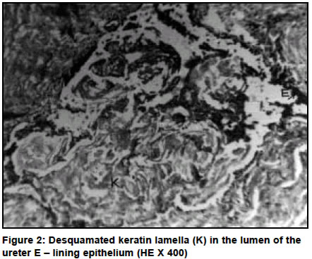
Paper Received: October 2002. Paper Accepted: February 2003. Source of Support: Nil. Code Number: is04025 ABSTRACT We report a case of a rare benign condition of the upper urinary tract histologically characterized by keratinising desquamative squamous metaplasia (KDSM). It is difficult to differentiate KDSM from other space occuping lesions of the upper urinary tract, but whenever diagnosis is made, conservative treatment can be employed. Key Words Keratinising, Desquamative squamous metaplasia, Upper urinary tract. How to cite this article: Nayak S, Puranik SC, Holla VV. Keratinising desquamative squamous metaplasia of the upper urinary tract: A case report. Indian J Surg 2004;66:105-6. INTRODUCTION The usual response of the transitional cell epithelium of the urinary tract to chronic irritation is proliferation. However in certain pathological conditions, squamous epithelium may be found replacing the transitional epithelium. We report a rare case of keratinising desquamative squamous metaplasia of the upper urinary tract presenting with obstructive pathology. CASE REPORT A 50-year-old man presented with right renal colic. He had a 2 year history of recurrent passage of multiple stones for which he had been operated. Investigations showed sterile pyuria and abundant acellular debris. Urea was 42 mg / dl and creatinine 1.8 mg/dl. Sonography revealed hydronephrosis of right kidney due to pelvic ureteric junction obstruction but no calculi. Findings on excretory urography of right kidney were hydronephrosis, dilated extrarenal pelvis, calculi in renal pelvis and mildly delayed excretory function. A right pyeloplasty was done. Multiple soft to firm brownish tissue bits were sent for histopathological examination. Microscopy revealed transitional epithelium focally replaced by squamous epithelium and overlined by laminated keratin (Figure 1). Also seen was a section of ureter with denuded keratin lamella in its lumen (Figure 2). The diagnosis given was keratinising desquamative squamous metaplasia of upper urinary tract. DISCUSSION Keratinising desquamative squamous metaplasia (KDSM) also referred to as leukoplakia or cholesteatoma of the upper urinary tract is an extremely uncommon disease.1-4 In this condition, the normal transitional epithelium of the urinary tract is replaced by squamous epithelium. Desquamation and accumulation of keratin debris leads to obstruction and symptoms of a space occupying lesion. As compared to upper urinary tract, squamous metaplasia is more commonly seen in the bladder where it is associated with squamous cell carcinoma related to bilharzial infection.2,5 In the upper urinary tract it is associated with urolithiasis and hydronephrosis as seen in the present case. Other associated conditions include recurrent urinary tract infection and tuberculosis.1,4,5 In few instances no associated pathology is present.1 Although the pathogenesis remains unclear, the possible explanations offered include (i) a reactive epithelial response, (ii) a congenital etiology or (iii) a spontaneous epithelial translation.1,3,4 The clinical diagnosis of KDSM is not easy. Recurrent renal colic, passage of desquamated keratinised cells and a lamellar configuration on excretory urography are suggestive of KDSM.1,4,5 The differential diagnosis includes urothelial tumor, radiolucent stone, blood clot, tuberculosis and papillary necrosis.1,5 Conservative surgery with follow up of patients has been justified by most authors since definite evidence of KDSM relationship with carcinoma is still lacking.1,3-5 Few authors however still consider it to be a premalignant condition.2,6 REFERENCES
© 2004 Indian Journal of Surgery. The following images related to this document are available:Photo images[is04025f1.jpg] [is04025f2.jpg] |
| |||||||||
