 |
| About Bioline | All Journals | Testimonials | Membership | News |
|
||||||
|
||||||
Indian Journal of Surgery, Vol. 66, No. 2, Mar-Apr, 2004, pp. 115-118 Case Report Necrotizing enterocolitis in adults: A study of four cases Avinash N. Katara, Vinod A. Chandiramani, Rajeev Soman,* Anita Bhaduri,** Devendra C. Desai*** Departments of Surgery, *Medicine, **Pathology and ***Gastroenterology, P.
D. Hinduja National Hospital and Medical Research Centre, Veer Savarkar Marg,
Mahim, Mumbai - 400016, India.
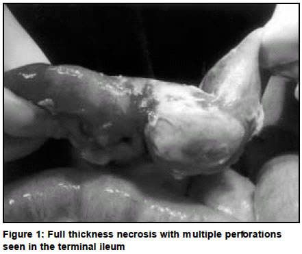
Paper Received: May 2003. Paper Accepted: August 2003. Source of Support: Nil. Code Number: is04031 ABSTRACT Necrotizing enterocolitis is an acute disease that primarily affects premature neonates of low birth weight, and has a very high morbidity and mortality. The incidence in adults is significantly less, with lower mortality rates. Of those who survive, many are left with complications related to short gut syndrome. We report 4 cases of necrotizing enterocolitis all of whom underwent early surgical intervention and had good recovery, followed by a review of the literature on the subject. Key Words Necrotizing enterocolitis, Adults. How to cite this article: Katara AN, Chandiramani VA, Soman R, Bhaduri A, Desai DC. Necrotizing enterocolitis in adults: A study of four cases. Indian J Surg 2004;66:115-8. INTRODUCTION Necrotizing enterocolitis is an acute disease that primarily affects premature neonates of low birth weight, and has a very high morbidity and mortality. The incidence in adults is significantly less, with lower mortality rates. The overall outcome depends on clinical staging and radiological and haematological parameters. Of those who survive, many are left with complications related to short gut syndrome. CASE REPORTS Case 1 A 35-year-old male presented with acute generalized abdominal pain, vomiting and fever of three days' duration. He was febrile and tachycardic. The abdomen was distended with minimal tenderness, and rectal examination was normal. Haematological investigations were normal except for a WBC count of 6,900/cu mm, and biochemical investigations showed a serum albumin of 1.8 g%. A plain abdominal X-ray showed free air under the diaphragm. The clinical picture with a normal leucocyte count did not fit into the classical features of perforative peritonitis, and so a CT scan abdomen was done. The CT identified moderate ascitis and stranding of fat planes around the caecum. Exploratory laparotomy revealed gross purulent peritonitis with three perforations in the thickened terminal ileum each about 2 to 3 mm in diameter (Figure 1). The caecum and ascending colon were congested and oedematous. A right hemicolectomy was done and the terminal ileum and transverse colon were brought out as separated stomas. He had an uneventful but prolonged postoperative course. Histopathology of the resected specimen showed extensive ulceration of the ascending and transverse colon and terminal ileum, of no specific aetiology. The continuity of bowel was restored six months later with an ileo-transverse anastomosis. Case 2 A 44-year-old male presented with clinical features suggestive of acute intestinal obstruction of seven days' duration and signs of peritonism. Apart from a high WBC count of 14,600/cu mm, haematological and biochemical investigations were normal. Radiological investigations confirmed the clinical impression of small bowel obstruction and cholelithiasis. Laparotomy showed gross peritonitis with perforation in gangrenous segments of the terminal ileum. Around three feet of the small bowel was resected and a primary anastomosis performed. Histopathology revealed a non-specific picture of infarction necrosis of the bowel wall without any evidence of major vessel thrombosis. Case 3 A 55-year-old male presented with abdominal pain of five days' duration and on examination was found to have signs of intestinal obstruction. His haematological and biochemical profiles were normal. A plain abdominal X-ray and CT scan confirmed the presence of bowel obstruction and the latter showed patent mesenteric vessels with haziness of the mesentery. Exploratory laparotomy revealed faecal peritonitis and a full thickness necrosis of the terminal ileum with perforation. A localized resection with primary anastomosis was undertaken. Histopathology showed ischaemic ulceration of the ileum covered by fibrinous and neutrophilic exudates, and ischaemia with transmural necrosis of the bowel. Case 4 A 48-year-old male presented with acute abdominal pain on the background of episodes suggestive of subacute bowel obstruction of over three months duration. Examination showed the patient to be tachycardic, in shock and the abdomen to be distended, guarded and rigid. Apart from a leucocytosis of 14,500/cu mm, other haematological and biochemical parameters were normal. A plain X-ray of the chest showed free gas under the diaphragm and abdominal X-ray showed dilated bowel loops. A CT scan confirmed these findings. A laparotomy showed gross faecal contamination of the peritoneal cavity with a perforation in the midst of a dusky terminal ileum. The caecum appeared congested and oedematous. The terminal three feet of ileum, caecum and appendix were resected and a terminal ileostomy was brought out. The ascending colon was closed in two layers. Histopathology revealed an ulcerated small bowel covered with granulation tissue, with mild inflammatory changes in the caecum. DISCUSSION Necrotizing enterocolitis (NEC) is an acute disease that primarily affects premature neonates of low birth weight. The incidence of NEC ranges from 0.3 to 2.4 / 1000 live births but is approximately 10% in infants weighing less than 1500 g.1,2 Although in adults the incidence of NEC is much lower and it has a lower mortality, it remains associated with considerable morbidity. NEC in adults is common in the developing countries and its aetiology is multifactorial. Infectious agents, inflammatory mediators and circulatory disturbance have all been implicated in the aetiology and pathogenesis of NEC. Epidemic outbreaks of NEC point to the role of infectious agents, especially when the same organism is isolated from stool, blood and peritoneal fluid. The common organisms implicated are bacteria like Klebsiella, E. Coli, Enterobacter, Pseudomonas, Clostridia and Staphylococcus epidermidis, viruses like Corona virus, Rota virus and Entero virus and rarely, fungi like Candida albicans.3 Enteropathogenic viruses are believed to infect epithelial cells resulting in cell destruction, necrosis and intestinal perforation. However, there are indications to suggest that infection is not mandatory for the development of NEC. It is also suggested that reduced mesenteric blood flow leads to ischaemia, which in turn causes hypoxic cell damage and release of inflammatory mediators. The re-perfusion of these tissues results in the release of oxygen free radicals that in turn cause loss of cellular integrity. The final common pathway in the development of NEC is believed to involve mediators like platelet-activating factor, endotoxin lipopolysaccharide, tumour necrosis factor and other cytokines, prostaglandins and leukotrines. Other factors believed to play a major role in the neonates and infants are prematurity, low birth weight, enteral feeding and immunological deficiencies, particularly of the IgA secretory component, intestinal T-lymphocytes coupled with a poor antibody response.3 Sporadic cases are associated with nutritional disorders, alcoholism, malabsorption and are seen after gastric or pancreatic resection.4 Raw peanuts, soyabean and sweet potatoes contain trypsin inhibitors which block intestinal proteases that could otherwise inactivate b-toxin produced by clostridium perfringens.4 In our study, we found a few factors common to all the patients. They were all smokers and consumed alcohol regularly. They all had a poor nutritional status and were from the same geographical area. All our patients were anorexic and had significant weight loss, which prompted the clinician to consider tuberculosis in the differential diagnosis. These patients present with a constellation of symptoms and signs that may vary from subtle food intolerance and change in bowel pattern to a catastrophic deterioration in general condition with severe abdominal distension and bloody stools. There can be gastrointestinal dysfunction in the form of abdominal distension, vomiting, bilious drainage from feeding tubes or bloody stools. Systemic illness is indicated by the presence of fever, dyspnoea, lethargy and hypotension. NEC can be clinically and radiologically staged into three stages, as proposed by Bell and coworkers5 and later modified by Walsh and Kliegman.6 Stage 1 (Suspected NEC)
Stage 2 (Definite NEC)
Stage 3 (Advanced NEC) Intestinal pneumatosis is pathognomonic of NEC. There could be other radiological features such as multiple dilated loops of the small bowel, fixed intestinal loop suggestive of localized ischaemia, diminished bowel gas due to poor motility, intrahepatic gas in the portal venous system, or pneumoperitoneum. No individual laboratory test is diagnostic though haematological analysis may show abnormally raised or reduced leucocyte counts with a shift towards immature precursors. The classical triad of increasing thrombocytopenia, acidosis and hyponatraemia suggests severe NEC, and these are the patients who are more likely to require surgical intervention. In our study group, all the patients had a history indicative of a prolonged disease process which did not relate with the signs. Examination suggested acute pathology, and all our patients were moribund and required emergency surgical intervention. Their white cell counts ranged from 6,000/cu mm to 13,000/cu mm. This discrepancy in the clinical and haematological picture warranted a CT scan of the abdomen in all the patients. Treatment is largely supportive and consists of bowel rest, gastrointestinal decompression, fluid resuscitation and antibiotic therapy. A few patients may require correction of hypotension, respiratory dysfunction, anaemia, coagulation disorders or acid-base imbalance. Surgical intervention is necessary if there is intestinal necrosis or frank perforation or when there is clinical deterioration over 1224 hours despite intensive medical support, as evidenced by persistent or worsening metabolic acidosis, increasing ventilatory requirement, deteriorating haematological parameters and persistent thrombocytopenia. At surgery, one of the four options may be resorted to: A) Resection of the necrotic bowel and primary anastomosis, which is the procedure of choice in patients who are stable and have only localized disease, B) Resection of the necrotic bowel and exteriorization of the ends as a proximal stoma and a distal mucous fistula. Continuity of the bowel may be restored weeks to months later after a radiological exclusion of a distal stricture, C) Proximal diverting jejunostomy done in the presence of extensive small bowel disease and D) Peritoneal drainage under local anaesthesia for decompression and release of contaminated material done as a palliative procedure in moribund patients. Complications in these patients could occur as a result of the primary disease process or due to the management. The immediate complications are intestinal perforation with intra-abdominal abscess and peritonitis, shock, sepsis, acute tubular necrosis and acute renal failure. The delayed complications include short bowel syndrome, malabsorption syndromes, recurrence, bowel stricture and atresia, enterocolic fistula, anastomotic leak and cholestasis. There could also be delayed systemic complications like chronic salt, potassium and water depletion and weight loss. The overall outcome depends on clinical staging and radiological and haematological parameters. The factors that point to a poor prognosis are A) Clinical: Signs of peritonitis, presence of perforation or pan-NEC, B) Systemic: Shock, presence of disseminated intravascular coagulation, acute renal failure, multi-system organ failure, or rapid progression of the disease, C) Investigational: Progressive thrombocytopenia, severe acidosis, electrolyte imbalance, progressively decreasing absolute neutrophil count and D) Radiological: Fixed intestinal loop, hepatic portal venous gas or perforation. REFERENCES
© 2004 Indian Journal of Surgery. The following images related to this document are available:Photo images[is04031f1.jpg] |
| |||||||||
