 |
| About Bioline | All Journals | Testimonials | Membership | News |
|
||||||
|
||||||
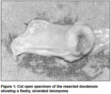
Indian Journal of Surgery, Vol. 66, No. 3, June, 2004, pp. 171-173 Case Report Pancreas-sparing duodenectomy for leiomyoma of the third part of duodenum: A case report Yadav TD, Sinha Surajit , Kaushik Robin Department of Surgery, Government Medical College and Hospital, Chandigarh-160030 Code Number: is04042 ABSTRACT Leiomyomas are benign neoplasms of smooth muscle that are rarely seen in the duodenum. They are usually asymptomatic and detected incidentally either on routine radiological investigations or at laparotomy or on autopsy. We present the case of a patient with leiomyoma arising from the third part of the duodenum who underwent pancreas-sparing duodenectomy for removal of the tumour.INTRODUCTION Leiomyomas are benign neoplasms of smooth muscles that commonly arise in tissues with a high content of smooth muscle such as the uterus and the stomach. Within the bowel, the commonest site of origin is the stomach. Duodenal leiomyomas are rare, benign tumours that are usually asymptomatic. When symptomatic, duodenal leiomyomas may present with abdominal pain or mass, obstruction or gastrointestinal bleeding.[1],[2],[3],[4] The treatment of duodenal leiomyomas has traditionally been duodenotomy, local tumour excision and primary duodenal closure, with pancreaticoduodenectomy reserved for large tumours, where there is a doubt of malignancy, or when the ampulla or pancreas are involved.[1],[2],[4] A case of leiomyoma of the third part of the duodenum who underwent pancreas-sparing duodenectomy (PSD) to remove the tumour is reported. CASE REPORT A 67 year old man presented with epigastric pain and recurrent hematemesis and malena over a period of one year. The pain occurred two to three hours after meals. Clinical examination was uneventful. Laboratory investigations were within normal limits. Barium meal examination showed a filling defect in the proximal third part of the duodenum. Endoscopy revealed mild oesophagitis with a slightly elevated, friable, pinkish ulcerated mass in the third part of the duodenum, biopsy of which revealed leiomyoma.The patient was taken up for surgery and a Type I b pancreas-sparing duodenectomy (see discussion) was performed after confirming the benign nature of the tumour by frozen section. The infra-ampullary duodenum was carefully dissected off the pancreas and the plane between the pancreas and duodenum developed by ligating the small vessels here. The duodenum was transected below the ampulla and removed alongwith the first few centimeters of the jejunum. The second part of the duodenum (infra-ampullary) and the jejunum were anastomosed as a duodenojejunostomy (end to end, hand sewn, single layer). Cut section of the resected duodenum showed a 2 cm by 1 cm fleshy, ulcerated mass arising in the third part of the duodenum [Figure - 1]. The patient developed bilious vomitings following return to oral feeds on the fourth postoperative day. Reinsertion of a nasogastric tube was followed by high daily output of upto 1500 ml per day. When this persisted beyond the seventh day, a barium meal follow through was done, but no mechanical cause for the obstruction could be visualized. The patient was put on prokinetics (Injection metoclopramide 10 mg, intravenous, thrice daily) and gradually his Ryle′s tube output reduced. Oral intake could be resumed by the eleventh post-operative day and he was discharged a day later. Presently he is well on a follow up of nearly three years. The histopathology report of the resected tissue revealed leiomyoma of the duodenum. DISCUSSION Pancreas-sparing duodenectomy (PSD) was first described in humans for the treatment of benign duodenal disease in 1995.[5] The procedure involves removal of the duodenum without removal of the adjacent pancreas, thereby avoiding unnecessary resection of the pancreas and also, the performance of bilio-and pancreatico-enteric anastomosis, each with its inherent problems. PSD has been reported in the treatment of infra-ampullary duodenal pathology such as simple adenomas, adenocarcinoma, and leiomyosarcoma.[5],[6],[7],[8],[9] Although technically demanding, the advantages are obvious when compared to the classical Whipple′s for malignant disease. The procedure shortens the operative time, reduces the blood loss, and avoids the need for pancreatico- and bilio-enteric anastomosis.[9] The advantage of PSD over simple tumour excision for a benign pathology lies in preventing recurrences by removing the affected duodenum, and by avoiding unnecessary resection of the adjacent pancreas. For adenomas, where upto 40 % may have carcinoma in situ, and field changes may be present throughout the duodenum, PSD may be curative, with a much lesser morbidity than a pancreaticoduodenectomy.[6] Depending upon the extent of resection, PSD is classified into Type I, II and III.[5] Type I PSD represents a subtotal duodenectomy that preserves the major and minor papillae. It has been further subdivided into I a (where resection of the duodenum is done above the papilla) and I b (where resection is done below the papilla). In Type II, the papilla is left as a button on the pancreatic head after a total duodenectomy, with reconstruction using technique of papillo-jejunostomy. In Type III, the terminal portions of the CBD and pancreatic duct are exposed and anastomosed to an isolated loop of jejunum. This patient continued to have a "functional" obstruction in the post-operative period that manifested as high Ryle′s tube output. We believe that this is a ′duodenal ileus′, the exact cause of which is not known. Although delayed gastric outlet obstruction occurring at a late stage has been reported previously,[6] we could not come across any report of ileus occurring immediately in the postoperative period. It may be possible that the procedure leads to a temporary compromise of the complex pancreatico-duodenal blood circulation, causing a temporary ischemia that manifests as local ileus. Another possibility is simply oedema of the anastomosis, but the exact cause of this "ileus" remains unclear. It has become our practice, however, to perform a feeding jejunostomy (Witzel technique) in addition to PSD to tide over this complication in the post-operative period. REFERENCES
Copyright 2004 - Indian Journal of Surgery The following images related to this document are available:Photo images[is04042f1.jpg] |
| |||||||||
