 |
| About Bioline | All Journals | Testimonials | Membership | News |
|
||||||
|
||||||
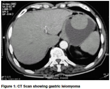
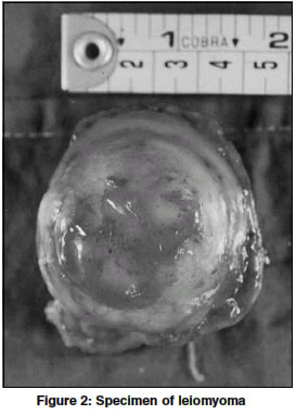
Indian Journal of Surgery, Vol. 66, No. 3, June, 2004, pp. 181 Images Gastric leiomyoma presenting as massive haematemesis Akolekar Deepika , Jaiswal Amit , Dharap Satish B Department of General Surgery, L. T. M. M. College and L. T. M. G. Hospital, Sion, Mumbai-400022 Code Number: is04047 A 75 year old chronic alcoholic male patient presented with massive haematemesis and malena. Upper GI endoscopy showed a sessile, vascular mass in the fundus of stomach with central umblication. CT scan abdomen however revealed a pedunculated tumour arising from fundus of stomach with high degree of vascularity suggestive of a stromal tumour. He underwent a laparotomy and wide excision of the 4 x 4 cm vascular, sessile mass (located on the anterior wall of the stomach at the junction of fundus and body) with one cm margin followed by primary closure of stomach in two layers. [Figure - 1] Histopathology revealed interlacing bundles of smooth muscle fibres in a characteristic whorl pattern with no mitotic activity suggestive of leiomyoma of stomach with no evidence of malignancy. Treatment modalities for such tumours include endoscopic polypectomy[1] which is possible in case of pedunculated tumours, wide local excision with primary repair and laparoscopic resection using Endo GIA staplers.[2] [Figure - 2] REFERENCES
Copyright 2004 - Indian Journal of Surgery The following images related to this document are available:Photo images[is04047f2.jpg] [is04047f1.jpg] |
| |||||||||
