 |
| About Bioline | All Journals | Testimonials | Membership | News |
|
||||||
|
||||||
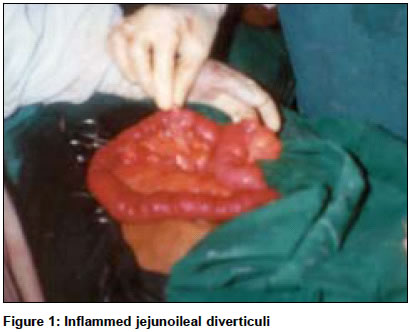
Indian Journal of Surgery, Vol. 66, No. 5, September-October, 2004, pp. 289-290 Case Report Primary acquired jejunoileal diverticulitis: A rare presentation Mohapatra Biswajit Department of Surgery, Vesaj Patel Hospital and Research Centre, Rourkela, Orissa Code Number: is04074 ABSTRACT Primary jejunoileal diverticulitis is a very rare disorder found only in 0.2 to 1.1% of the adult population. The incidence increases with age, peaking in the sixth and seventh decades. In this case report I present a 72-year-old patient who developed intestinal obstruction due to jejunoileal diverticulitis.KEY WORDS: Jejunoileal diverticulitis, Obstruction INTRODUCTION Small bowel diverticulitis occurs less frequently than large bowel diverticulitis. Acquired jejunoileal diverticulas are uncommon and asymptomatic in the majority of patients. Chow et al have suggested that over 90% patients with jejunoileal diverticula may manifest non-specific symptoms. Serious complications of jejunoileal diverticulitis include massive haemorrhage, small bowl obstruction, volvulus, perforation, and sepsis. Because of its delayed presentation and diagnosis it carries a very high reported mortality of 25 to 30%, considering most of these patients are elderly and have multiple co-morbid diseases.[1] CASE REPORT A 72-year-old patient underwent Freyer′s prostadectomy for benign prostatic hyperplasia. The operation was uneventful. The patient developed high-grade fever with rigor and chills after 6 hours of operation and was treated with dexamethasone 8 mg.stat and cold sponging. The total count was 16000/ c mm on investigation. The patient was on Ceftriaxone, Amikacine and Metronidazole. The patient passed flatus on the 2nd day and oral liquid was started. But the patient started complaining of abdominal pain and stopped passing flatus on the 3rd evening. The patient was treated conservatively for the next 3 days without any improvement. X-ray abdomen standing view showed multiple air fluids levels, suggestive of established intestinal obstruction. On the 7th postoperative day exploratory laparotomy was done to relieve the obstruction. Multiple inflamed acquired jejunoileal diverticula were found on the mesenteric border [Figure - 1]. Obstruction was due to adhesion of one of the inflamed jejunal diverticulums with the distal ileal mesentery, causing an acute bent. Simple adhesiolysis was done and the abdomen was closed with drains. Patient′s recovery was smooth and was discharged on the 8th day of the second operation. DISCUSSION This case study allows for a review of primary acquired jejunoileal diverticula causing intestinal obstruction. They are formed by herniation of mucosa and submucosa through the muscular wall of the intestinal wall. They are always multiple and occur on the mesenteric border. They are technically pseudodiverticula as they lack the muscular coat. Their size varies from a few millimetres to more than 10 cm.[2] They tend to be larger and higher in number in the proximal jejunum and they are fewer in number and smaller in size towards the distal ileum. Sixty per cent of patients with jejunoileal diverticulosis are asymptomatic. Thirty per cent develop a symptom complex described by Edwards as flatulent dyspepsia, consisting of epigastric pain, abdominal discomfort, and flatulence an hour after a meal.[3] The remaining 10% develop complications requiring surgery. Two main radiological methods to diagnose jejunoileal diverticulitis are enteroclysis and contrast enhanced C.T. scan of abdomen.[2] During enteroclysis a water-soluble contrast medium is injected into the proximal duodenum through a tube. Radiological finding includes saccular outpouching at the mesenteric border of the small intestine. The CT scan findings generally demonstrate an inflammatory mass adjacent to the small bowel loop. Small air bubbles within the mass can be seen. Mechanical intestinal obstruction occurs in 2.3 to 4.6% of cases of jejunoileal diverticulitis.[4] This may be the result of pressure on the intestinal wall from distended diverticula, stricture or adhesion from recent or past diverticulitis, intussusceptions at the site of diverticulum, enteroliths developed within the diverticula, or volvulus of the diverticula-containing segment. The involved segment with its diverticula filled with fluid is heavier then the non-involved part. This loaded segment swings like a pendulum to initiate the volvulus. In my case adhesion of the inflamed diverticuli to the adjacent mesentery causing intestinal obstruction. The management of primary jejunoileal diverticulitis is quite variable, depending on the presenting symptoms. Initial management is always a conservative approach with antibiotics and bowel rest. The definitive management of perforated diverticulitis is surgical resection of the small bowel segment. In cases of intestinal obstruction not responding to conservative management, laparotomy is indicated. However, simple diverticulectomy is strongly discouraged in the literature due to a very high rate of leakage. Because of the mesenteric location of these diverticula, simple diverticulectomy may impair blood flow and lead to anastomotic leakage.[1] The surgical therapy of obstruction due to volvulus consists of simple untwisting or resection of the involved segment. Sometimes simple adhesiolysis is needed to relieve the obstruction, which was done in my patient. Cross and Synders[5] reported the use of laparoscopically directed small bowel resection for jejunoileal diverticulitis with perforation. The laparoscope was utilised to diagnose the disease and to run the small bowel. A 5-cm incision was given directly over the pathology and the involved segment was exteriorised to do the resection and extra corporeal anastomosis. In conclusion, jejunoileal diverticulitis should not be regarded as an insignificant entity. It is essential to include the jejunoileal diverticula in the differential diagnosis of abdominal pain in patients of the 6th and 7th decades, as a delayed diagnosis increases the mortality rate significantly in these groups. Liberal use of C.T. scans for the work-up of abdominal pain will limit the number of missed diagnoses. REFERENCES
Copyright 2004 - Indian Journal of Surgery The following images related to this document are available:Photo images[is04074f1.jpg] |
| |||||||||
