 |
| About Bioline | All Journals | Testimonials | Membership | News |
|
||||||
|
||||||
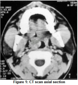
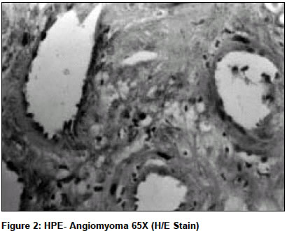
Indian Journal of Surgery, Vol. 66, No. 5, September-October, 2004, pp. 293-294 Case Report Angiomyoma of soft palate - A case report Srinath VS, Meher Ravi, Sabherwal Anup, Sharma Naveen Departments of Pathology, Maulana Azad Medical College and associated Lok Nayak, G. B. Pant Hospitals and G.N.E.C. New Delhi - 110002 Code Number: is04076 ABSTRACT Angiomyomas are benign neoplasm thought to originate from vascular smooth muscle. They have a propensity to arise from the GIT and female genital tract (uterus) and subcutaneous tissue. The oral cavity is uncommon site for angiomyoma. Here is an interesting case report of a rare palatal angiomyoma. A brief review of the literature and histological variations have also been described.KEY WORDS: Angiomyoma, soft palate CASE REPORT A 35-year-old male presented to the ENT OPD with a swelling of his soft palate, which was gradually increasing in size for the past one year. It was not associated with pain, bleeding, dysphagia or odynophagia. There were no dental complaints and there were no symptoms suggestive of any local infection. On examination there was an intra-oral mass averaging 2´3 cm, arising from the right soft palate extending over to the hard palate with an intact mucosal lining. It was firm on palpation and was non-pulsatile. A FNAC of the swelling was performed intra-orally, which yielded only blood. A contrast enhanced CT scan [Figure - 1] revealed an enhancing soft tissue mass arising from the right soft palate, without bony erosion. A provisional diagnosis of hemangioma was made. The patient was taken up for excisional biopsy of the intra-oral mass under general anaesthesia via transoral approach. A histopathological analysis [Figure - 2] of the specimen revealed an angiomyoma. DISCUSSION A total of 139 cases of leiomyomas of the oral cavity and pharynx have been reported till date (Hatch, Davis et al, 2001),[1] out of which only 19 have been palatal angiomyomas (Svane, Smith, Cosentino, 1986).[2] Angiomyomas are leiomyomas of vascular smooth muscle origin.[3],[4] Benign smooth muscle neoplasm have been classified into[5]
Myxoid angiomyoma is a rare variant of angiomyoma (Holder, Dellinger, 2001).[6] A review of the literature showed that the mean age of presentation of oral leiomyoma is 41 years, with male sex predominance (54%). The most common sites of presentation were lips (27%), followed by tongue (18.3%), cheek and palate (15.49%), gingival (8.45%) and mandible (5.63%) (Baden, Doyle, Lederman, 1994). Oral leiomyomas are believed to be derived from vascular smooth muscle and the division between angiomyoma and leiomyoma is based on the degree of vascularity. Histological differentiation must be made from neurofibroma, other spindle cell tumours, myofibroma, and granular cell tumours and leiomysarcomas. In case of histological controversy, immunohistochemical markers like muscle specific actin, desmin, myoglobin, S-100 protein, vimentin can be used. Our case had both smooth muscles and vascular proliferates. Smooth muscle cells had blunt cigar-shaped uniform nuclei with no demonstrable atypia or mitotic activity. Although routine H/E and cytochemical (VGE-Von Grieson elastin) staining was sufficient for diagnosis, we put up an antibody panel consisting of smooth muscle actin, desmin and factor VIII. Immunohistochemistry was done using standard ABC method after antigen retrieval. The lesion was positive for all three markers, though desmin showed only focal positivity. We conclude that angiomyomas are very rare tumours, which are benign by nature. They should be differentiated from neurofibroma, other spindle cell tumours, myofibroma, granular cell tumour and malignant leiomyosarcoma. Immunohistochemistry is precise and reliable for the definitive diagnosis of an angiomyoma. The prognosis of oral leiomyoma is excellent after complete excision. REFERENCES
Copyright 2004 - Indian Journal of Surgery The following images related to this document are available:Photo images[is04076f1.jpg] [is04076f2.jpg] |
| |||||||||
