 |
| About Bioline | All Journals | Testimonials | Membership | News |
|
||||||
|
||||||
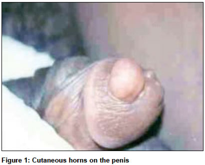
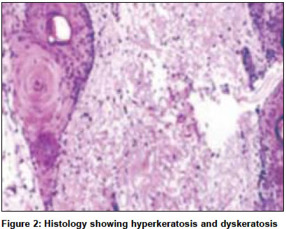
Indian Journal of Surgery, Vol. 66, No. 5, September-October, 2004, pp. 296-297 Case Report Cornu cutaneum-cutaneous horn on the penis Rekha A, Ravi A Department of Surgery, Sri Ramachandra Medical college and research institute, Porur, Chennai Code Number: is04078 ABSTRACT We report a 38-year-old man presenting with a penile cutaneous horn. Cutaneous horns are seen in sun-exposed areas and their occurrence on the penis is uncommon. The association with malignancy on the penis makes proper identification of these lesions essential. Standard treatment involves local excision, but the presence of malignancy mandates a partial penectomy.KEY WORDS: Cornu cutaneum, penile horn, malignancy INTRODUCTION Cornu cutaneum (cutaneous horn) refers to a well-defined cone-shaped lesion with hyper-keratotic features. These are found most frequently on exposed skin.[1] Cutaneous horns occur only rarely on the penis. As up to a third of penile cutaneous horns are associated with underlying malignancy, early excision is advocated.[2] CASE REPORT A 38-year-old man sought attention for nodules on his penis. He had no pain, itching or discharge. Examination revealed two nodules on his glans penis - the larger one was 1cm x1 cm and the smaller about 0.25cm x 0.25cm in size [Figure - 1]. There was no inguinal lymphadenopathy. Both nodules were excised with a rim of normal tissue and primary closure was achieved. Postoperative recovery was uneventful. Histopathology of the nodules revealed extreme hyperkeratosis, dyskeratosis, and acanthosis [Figure - 2]. Although there was no focus of malignancy there were a few areas showing squamous atypia. DISCUSSION Cutaneous horns are only rarely seen in areas not exposed to sunlight. Various lesions seen at the base of a cutaneous horn include squamous cell carcinoma, actinic keratosis, keratoacanthoma, Bowen′s disease, seborrheic keratosis, basal cell carcinoma, hemangioma, keratotic and micaeous pseudopapillomatous balanitis, Kaposi′s sarcoma, sebaceous adenoma and Paget′s disease of the female breast.[3] The first case of a cutaneous horn was described in 1854, and since then fewer than 100 cases have been reported.[4] The aetiology of penile horns is uncertain, although they are often found in association with warts, phimosis, naevi and in areas afflicted by trauma.[5],[6] Malignant change should be suspected in a rapidly growing lesion. Microscopically, a cutaneous horn shows marked hyperkeratosis, acanthosis, dyskeratosis, papillomatosis and chronic inflammatory infiltration of the adjacent dermis.[1] Malignant change has been reported in 12 out of the 100 cases.[7] In contrast, another study observed that while rapid growth is common, malignant degeneration is infrequent. There has been one case report of multiple penile horns.[8] Treatment options include wide surgical excision with careful histological examination to exclude a focus of malignancy. If malignancy is present in a penile cutaneous horn, the treatment involves partial penectomy with or without regional lymph node dissection. Therapy with carbon dioxide or Neodymium YAG laser is used for patients who refuse surgery.[2] While preliminary studies with laser are encouraging, partial penectomy remains the gold standard. REFERENCES
Copyright 2004 - Indian Journal of Surgery The following images related to this document are available:Photo images[is04078f2.jpg] [is04078f1.jpg] |
| |||||||||
