 |
| About Bioline | All Journals | Testimonials | Membership | News |
|
||||||
|
||||||
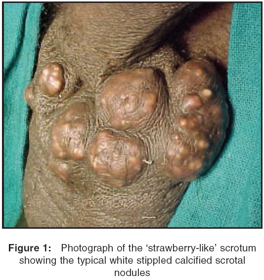
Indian Journal of Surgery, Vol. 67, No. 3, May-June, 2005, pp. 159 Images in Surgery Strawberry scrotum Singh Iqbal, Singh Nain, Sharma Naveen, Agarwal Sarla, Tandon R Departments of Surgery and Pathology, University College of Medical
Sciences (University of Delhi) & GTB Hospital, Delhi, India Code Number: is05045 Keywords: Calcified epidermal cysts, calcinosis cutis, scrotal calcinosis A 35-year-old man presented with painful scrotal swellings for the last 8 years and local itching with occasional discharge of whitish chalky material. Local examination revealed multiple diffuse nodular swellings [Figure - 1] about 6 x 8 cm, arising from the anterior scrotal skin covered with multiple white, hard, stippled chalk-like excrescences (strawberry). The external genitalia and groin did not show any abnormality. Under local anesthesia, he underwent en-bloc excision of the anterior scrotal skin and primary skin closure. Histopathology revealed scrotal dermal calcinosis with areas of dystrophic calcification, epitheloid cells, foreign body giant cells, sclerosis, and partial coalescence of calcified nodules without any inflammatory reaction. Scrotal calcinosis (SC) or calcinosis cutis is the presence of calcified masses within the scrotal skin or dermis. Though commonly idiopathic[1] recent reports suggest that the basic underlying abnormality may be inflammation and or infection of the scrotal epidermal inclusion cysts,[2] necrosis may serve as a nidus for dystrophic calcification of these cysts. The presence of a high content of calcium and phosphorus crystals in these calcified nodule suggests a degenerative origin and 'calciphylaxis' (deposition of calcium in the small blood vessels with ischemic necrosis and infection of the overlying skin) of the scrotal skin.[3] Due to the absence of subcutaneous fat in the scrotal skin, these calcifications were easily seen through the thin parchment-like thermosensitive scrotal skin. Superadded scrotal itching and infection may have contributed to cuticular necrosis causing the chalky deposits to be leached out and giving rise to its atypical 'strawberry-like appearance' and this may have contributed to the histological evidence of occasional areas of foreign body giant cell reaction in the absence of any inflammation, as seen in this case. References
Copyright 2005 - Indian Journal of Surgery The following images related to this document are available:Photo images[is05045f1.jpg] |
| |||||||||
