 |
| About Bioline | All Journals | Testimonials | Membership | News |
|
||||||
|
||||||
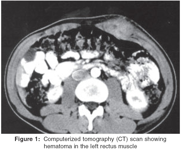
Indian Journal of Surgery, Vol. 67, No. 3, May-June, 2005, pp. 160-161 Images in Surgical Radiology Rectus sheath hematoma Gupta Sanjay, Kaushik Robin, Sharma Rajeev Department of Surgery, Government Medical College and Hospital, Chandigarh, India Code Number: is05046 Rectus sheath hematoma is a well documented but unusual cause of a painful abdominal lump that occurs as a result of accumulation of blood in the sheath of the rectus abdominis muscle, either secondary to rupture of the epigastric vessels, or due to tear of the muscle.[1] It may be misdiagnosed, leading to the performance of unnecessary surgery. Such a case is reported and the condition briefly reviewed. A 25-year-old cigarette vendor presented to the emergency with increasing swelling and pain in the left hypochondrium, just lateral to the midline for duration of 7 days. There was no fever or history of trauma. General examination revealed a tender mass, about 10 x 7 cm in size, just below the left costal margin, lateral to the midline. It did not disappear on lifting the head and shoulders of the patient. Systemic examination was unremarkable, and his routine hematological and biochemical investigations were all within normal limits. An ultrasound was asked for, but since it was inconclusive, a computerized tomogram (CT) scan was ordered [Figure - 1]. It revealed the mass confined to the upper part of the left rectus muscle, with minimal leak of contract within. A diagnosis of rectus sheath hematoma was made, and the patient put on analgesics and complete bed rest. We repeatedly asked the patient leading questions for any history of trauma, lifting of heavy weights or coughing and straining, but were unable to elicit any such details. The hematoma reduced in size over the next few days, following which the patient was discharged. Rectus sheath hematoma is often misdiagnosed. It occurs more often in women than in men (ratio 2: 1 to 3: 1), possibly as a result of the differences in muscle mass and the changes associated with pregnancy, and usually occur in the age group of 50-60 years.[1],[2] Usually, it occurs below the umbilicus, and is unilateral.[2],[3] Although the occurrence of rectus sheath hematoma has occasionally been reported to be spontaneous, predisposing conditions are usually demonstrable. These include external trauma, anticoagulation therapy, strenuous activity, coughing, lifting, sneezing, vomiting, pregnancy, blood dyscrasias, previous surgery, laparoscopic trocar injury, or injection of drugs. [1],[2],[3],[4] The diagnosis of the condition can be elusive, as it can mimic acute abdomen, and the patient may even develop shock because of the blood loss. This assumes importance in the setting of trauma, where the patient may undergo an unnecessary surgery with the diagnosis of intra-abdominal trauma, especially in centers where the facilities for imaging (ultrasound or CT scan) may not be adequate. Adequate clinical examination, and awareness of the fact that the mass does not move with respiration, and becomes fixed when the patient lifts the head while supine (Fothergill sign) may indicate the diagnosis[2] and surgery could be avoided. Conservative treatment is favored if the patient is hemodynamically stable and the hematoma is nonexpanding. If the hematoma has occurred secondary to anticoagulation, or has caused hemodynamic compromise, or has expanded on conservative management, the treatment is surgical, with evacuation of the hematoma and ligation of the bleeding vessel.[2],[3] References
Copyright 2005 - Indian Journal of Surgery The following images related to this document are available:Photo images[is05046f1.jpg] |
| |||||||||
