 |
| About Bioline | All Journals | Testimonials | Membership | News |
|
||||||
|
||||||
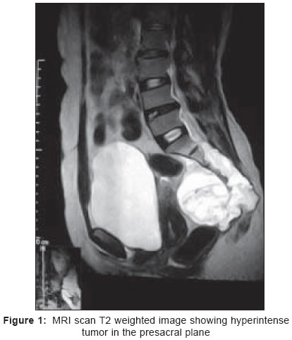
Indian Journal of Surgery, Vol. 67, No. 4, July-August, 2005, pp. 207-209 Case Reports Sacral chordoma - a report of two cases S. Rao BS, Menezes LT, Rao AD, John SK. Department of General Surgery, Fr. Muller Medical College Hospital, Kankanady, Mangalore, Karnataka Code Number: is05063 Abstract Chordoma is a rare, slow growing but locally aggressive malignant tumor derived from primitive notochordal elements, and it is usually found in the sacrococcygeal area. Chordomas are difficult to excise completely because preservation of sacral stability and sacral nerve pathways to the rectum and bladder limit the extent of surgery. The role of adjuvant treatment is uncertain and surgery remains the mainstay of its treatment. We present two such cases of sacral chordomas managed in our hospital. The clinical implications of these rare tumors are discussed.Keywords: Chordoma, Sacrococcygeal Chordomas are rare, slow growing but locally aggressive neoplasms derived from primitive notochordal elements. They are mainly found in the spheno-occipital and sacrococcygeal region.[1] Although, metastasis is infrequent at presentation, the prognosis for patients with chordoma of sacrum is reported to be poor and attributable in most cases to intralesional resection. The value of adjuvant treatment is uncertain and resection remains the primary mode of treatment. Chordomas are difficult to excise completely because preservation of sacral stability and sacral nerve pathways to the rectum and bladder limit the extent of surgery. Case reports Case 1 Case 2 Discussion Chordomas are neoplasms localized in the midline of the body with approximately 50% in the sacrococcygeal, 35% in the cervical spine and skull, and 15% in the thoracic spine and elsewhere. Male to female ratio is 3 : 1 and it is uncommon in individuals less than 40 years of age. Although the biological behavior of the chordomas is highly variable, most conventional chordomas are slow growing.[2] A sacral chordoma has presacral, subperiosteal extension, and also extension into the sacral canal. The sacrum and coccyx may be destroyed owing to tumor invasion. The rectum, bladder, uterus, and adnexae are either displaced or completely surrounded by the tumor. They are firm, grayish tumors, located and well encapsulated in soft tissue with variegated consistency and areas of hemorrhage. Metastases occur in only 10% of cases to lungs, liver, lymph nodes, skin, and muscles. Microscopically, it consists of pleomorphic cells in clusters amidst myxoid matrix. Immunohistochemically these cells are reactive to both cartilaginous and epithelial markers.[3] The presenting symptom in the most patients is local pain. About one-third of the patients also have radiculopathy due to irritation of the sciatic nerve or ileolumbar trunk.[4] Radiological examinations show soft tissue mass in the presacral region with expansion of the sacrum and irregular areas of bone destruction, which are well observed on CT scan or MRI. Chordoma can be suspected if a midline sacrococygeal tumor shows reduced uptake on bone scintigraphy with Tc-99m HMDP unlike most other bone tumors. Endorectal sonography can be used for diagnosis and guided biopsy.[5] Small posterior incision or trocar biopsy is recommended. Transrectal or transvaginal biopsy violates the containing membranes of the presacral fascia and periosteum and should be avoided to prevent tumor seeding.[9] Definitive treatment is by wide resection with normal tissue margins and avoidance of spillage. Kaiser et al.[6] reported that local recurrence rates correlated significantly with violation of tumor margins at initial surgery. En bloc sacral resection below the sacroiliac joint is a relatively straightforward procedure performed via a combined posterior-transperineal exposure. Tensionless wound closure may necessitate mobilization of soft tissue flaps but generally can be achieved without resorting to more elaborate reconstructive measures. In large tumors, a combined abdomino-sacral approach offers best results.[7] Radical excision can be done with limited postoperative morbidity and preservation of neurological function, including sphincter control, provided that one S2 nerve root is left intact.[8] Postoperative irradiation is advocated but its efficacy is a subject of further debate. Samson et al.[9] have advocated the use of radiotherapy to allow the surgeon to perform a marginal resection in case of high-sacral tumors allowing the preservation of nerve roots. Chemotherapy is of little value in these tumors. References
Copyright 2005 - Indian Journal of Surgery The following images related to this document are available:Photo images[is05063f2.jpg] [is05063f1.jpg] |
| |||||||||
