 |
| About Bioline | All Journals | Testimonials | Membership | News |
|
||||||
|
||||||
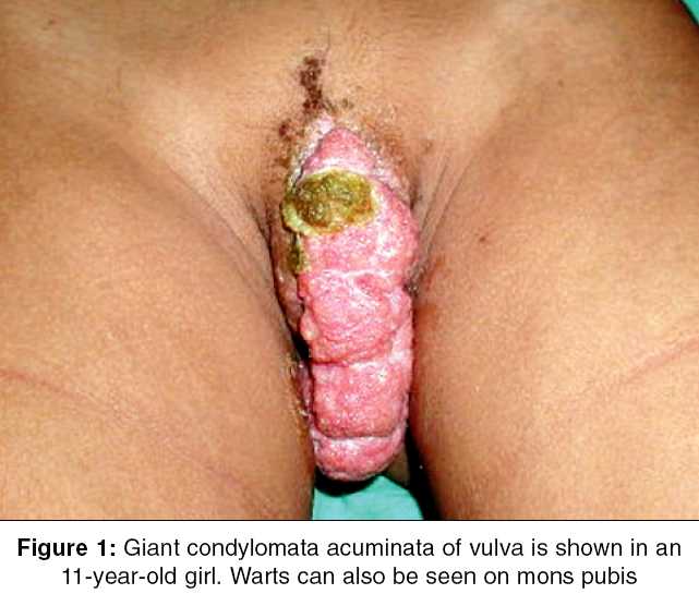
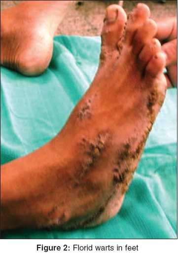
Indian Journal of Surgery, Vol. 67, No. 5, September-October, 2005, pp. 278-279 Images in Surgery Giant condylomata acuminata vulva in an adolescent girl Tewari M, Shukla HS Department of Surgical Oncology, Institute of Medical Sciences, Banaras Hindu University, Varanasi, Uttar Pradesh Code Number: is05087 An 11-year-old unmarried girl presented to us with a history of warty growths all over her body since birth. She denied any history of contact or sexual assault. Examination revealed an 8 x 2.5 cm fleshy mass arising from right half of her labia minora and majora entirely covering the vestibule [Figure - 1]. Vaginal mucosa appeared normal with an intact hymen. In addition, numerous cutaneous warts were present on her entire body surface and especially clustered on her feet [Figure - 2]. The VDRL and HIV antibody tests were negative. Examination of both the parents was normal. Patient′s mother Pap smear test revealed no abnormality. Bloodless excision of condylomata acuminata (CA) was done using the neodymium: yttrium-aluminium-garnet (Nd-YAG) laser under general anesthesia. The warts on her limbs were treated with Nd-YAG laser in two sessions at three weekly intervals. Histopathology suggested presence of HPV with no malignant change in the lesion. Polymerase chain reaction showed the presence of HPV type 11 in the genital CA. Warts result from infection with the double-stranded DNA virus, HPV, of which over 100 subtypes are now recognized.[1],[2] HPV infection is a sexually transmitted disease in adults. The modes of viral transmission in children remain controversial. These include perinatal transmission, auto- and hetero-inoculation, sexual abuse, indirect transmission via fomites, etc. The newborn babies can be exposed to cervical HPV infection of the mother during delivery. In utero transmission to the fetus may occur hematogenously, by semen fertilization, or as an ascending infection in the mother.[2] The genital warts initially are usually small finely branched structures with a narrow stalk and later may coalesce to form large cauliflower masses with broad dermal attachments, as was seen in our patient. Although, the vulva (particularly the vestibule, and labial folds) and the perianal skin are the sites most frequently involved, lesions may occur within the vagina, cervix or mons pubis in females. In males, these are typically located around the coronal sulcus, on the glans and the frenulum, at the meatus and sometimes on the shaft and surrounding skin. The rectum, anal canal and perianal areas can also be involved, particularly in homosexual men (but heterosexual men can also be affected). Only rarely do they turn malignant. These are thought to be a co-factor in the development of cervical cancer, although subject to much debate. Diagnosis is generally clinical. Biopsy is required only rarely. Condylomata acuminata is a therapeutic challenge. Cryotherapy (preferably with liquid nitrogen) is the preferred treatment. Trichloroacetic acid although widely used has unpleasant side effects. Patient applied treatments imiquimod and podophyllotoxin are the newer choices. Other modalities in resistant cases include electrotherapy, laser and surgical excision.[1],[3] Various types of lasers viz. Nd-YAG,[4] CO2,[5] pulsed dye[6] laser have been used either alone or with surgery with good clearance rates. Vulvar edema, bleeding and vulvodynia may occur following these treatments. Therapy is best administered in several applications at weekly intervals. Our patient responded well to Nd-YAG laser ablation of the warts and had no postoperative complications. References
Copyright 2005 - Indian Journal of Surgery The following images related to this document are available:Photo images[is05087f2.jpg] [is05087f1.jpg] |
| |||||||||
