 |
| About Bioline | All Journals | Testimonials | Membership | News |
|
||||||
|
||||||
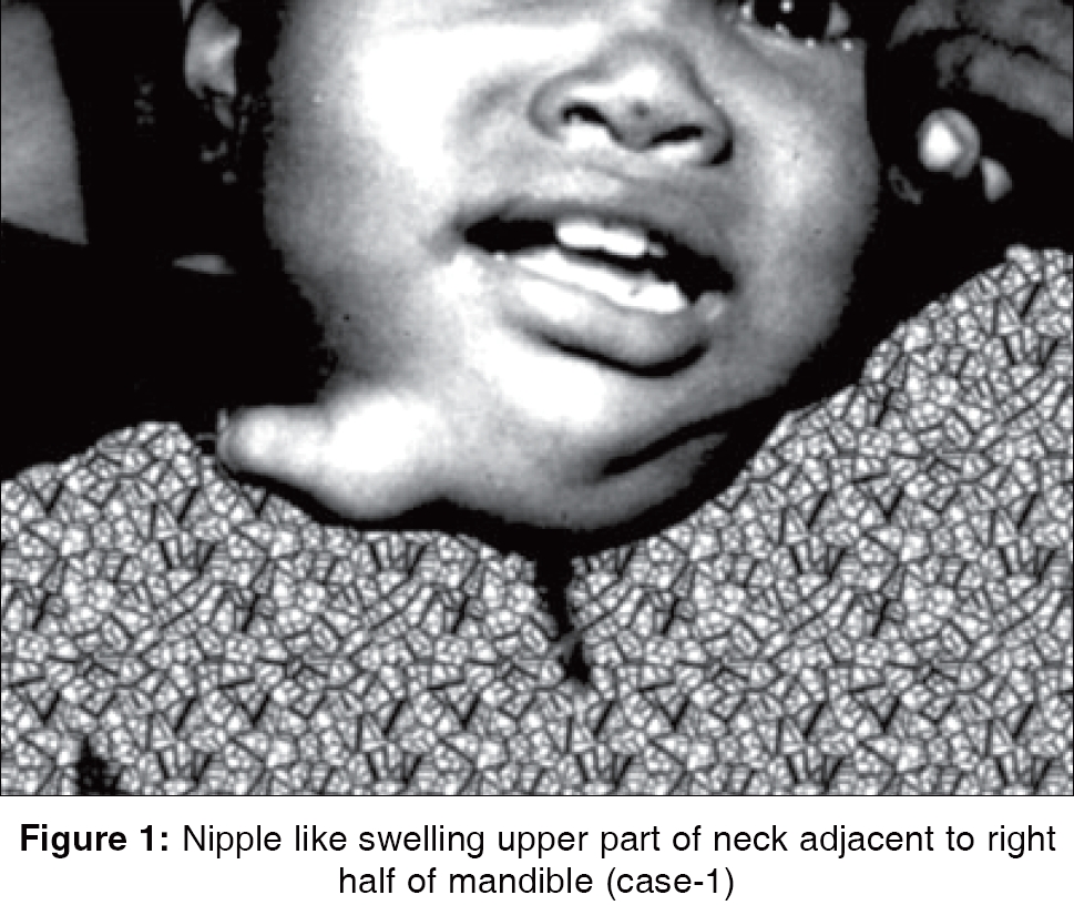
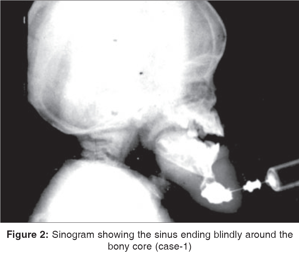
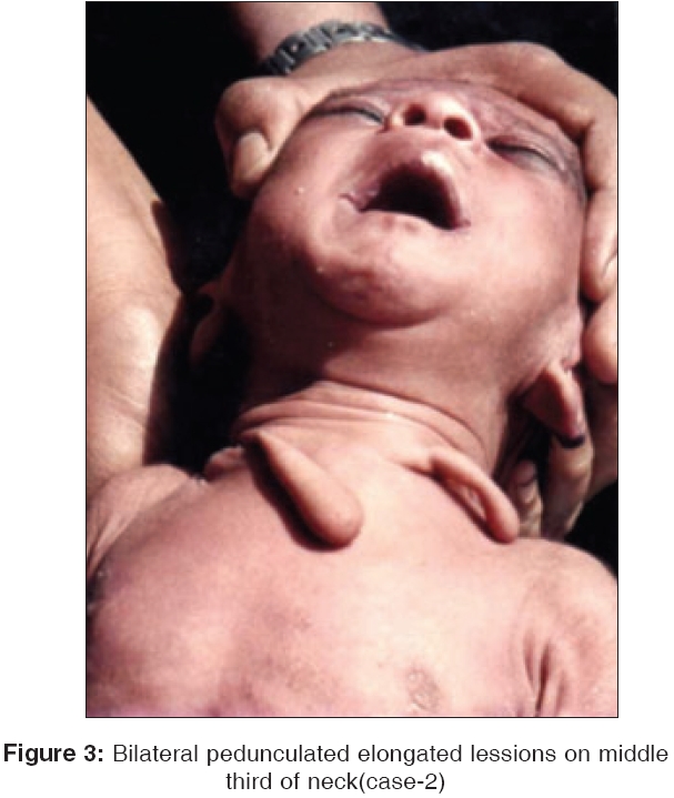
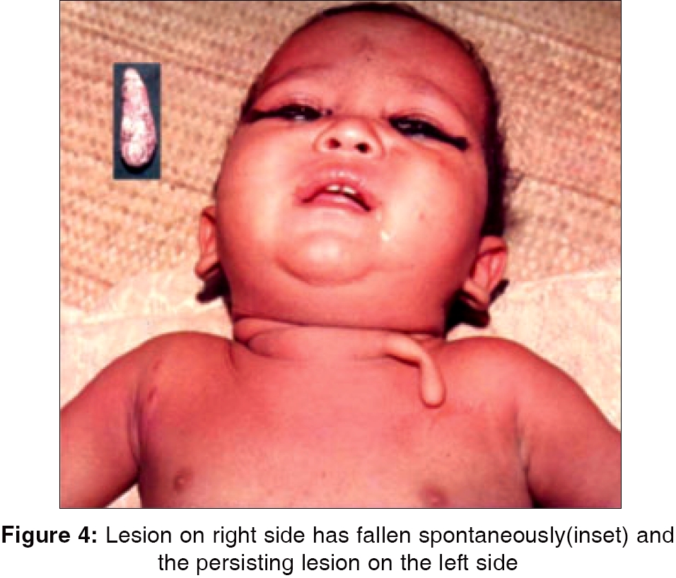
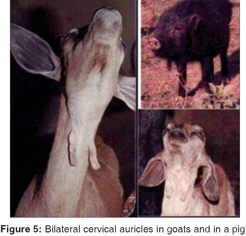
Indian Journal of Surgery, Vol. 68, No. 1, January-February, 2006, pp. 35-37 Case Report Cervical auricles from first and second branchial arches Maiti Sukumar Department of Surgery, Bankura Sammilani Medical College, Bankura, West Bengal Code Number: is06008 Abstract Cervical auricles were observed in three children. Operation (excision) could be done in two of the children. The anomalous auricle from first branchial arch contained an incomplete "cervico-auricular" sinus along with partial facial nerve palsy. Cervical auricles were also observed in a number of goats and in a pig. Morphological similarity was there in respect of location. Hereditary history was noted in the animals with cervical auricles.Keywords: Cervical auricles, first and second branchial arches, first branchial cleft sinus, anomalies in animals, family history. Introduction Inordinate and superfluous growths of the mesoderm components of the branchial arches are rare and may produce auricle like swellings in neck. Sinuses may be associated with the anomalies of branchial arches. Cervical auricles are very commonly found in goats and sometimes in other mammals. Case 1 Operation - a transverse incision encircling the swelling was made. Attached muscles were separated from the surface of the bony component, which was then divided from the mandible. The sinus tract was found ending around the distal surface of the bony core and the tract could not be traced further behind. Histological examination showed stratified squamous epithelial lining of the sinus tract. In three years′follow up the sinus or the auricle did not recur. Case 2 Case 3 In Goats In a Pig Discussion The rare and interesting facts worth mentioning in the first case are (1) First branchial cleft sinus opened on the top of an elevated hillock like lesion rather than on a flat surface. (2) Associated partial facial nerve palsy involving the adjacent branch (marginal mandibular) of facial nerve. When the baby cries the opposite angle of mouth is drawn to the left side (3) The cleft sinus was not extending up to the ear. In the usual cases the sinus/fistula extends from external auditory meatus to open just below the middle of the ipsilateral half of the mandible as ′Cervico-auricular′ fistula. (4) A bony core was present inside the anomalous structure. No such case with all the above features was found on the search of medical literature. The cervical auricles observed in goats, pigs and in the second patient were bilateral.[1] The position of the auricles in them indicated that the second branchial arch mesodermal element (hyoid sheath) was their source of development. The second arch defects are more common than any other arch anomalies. In the second and third cases of cervical auricles in the middle third of neck, of the present study as well as in none of the previously reported cases there had been any associated external fistula adjacent to or within the substance of cervical auricle. In some of the cases a cord of fibrous tissue could be traced upwards a few centimeters along the medial border of the sternomastoid muscle, but it never led up to the pharynx.[1] These lesions are objectionable from the cosmetic standpoint. The treatment of cervical auricles derived from second branchial arch is simple, as fistulae are not connected with the lesions.[1],[2] The mass can be excised with an elliptical piece of skin, a little bit of subcutaneous tissue and the underlying cartilage if any.[1],[2] Anomalies in the form of sinuses or cysts are more common than cervical tabs or auricles. Bilateral incidence of branchial anomalies may occur in 10-15% of patients.[3] There may be a positive family history in 10% of cases.[4] In ancient statues of Faunas (God of fertility and agriculture in Roman mythology) and Satyrs (The Greek equivalent of Faunas), cervical auricles were sometimes found.[5] The neck-ears of the statues were not modeled after the human ear, but looked like the cervical-ears of the goats. Cervical ears may also be found in the horses, pigs, sheeps and other mammals.[5] In the present series of cervical auricles, it was observed that there was no family history of the presence of the cervical auricles in the human cases but in cases of many of the affected animals (goats and pig) hereditary history of cervical auricles was present.References
Copyright 2006 - Indian Journal of Surgery The following images related to this document are available:Photo images[is06008f4.jpg] [is06008f5.jpg] [is06008f1.jpg] [is06008f2.jpg] [is06008f3.jpg] |
| |||||||||
