 |
| About Bioline | All Journals | Testimonials | Membership | News |
|
||||||
|
||||||
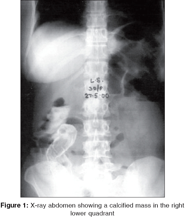
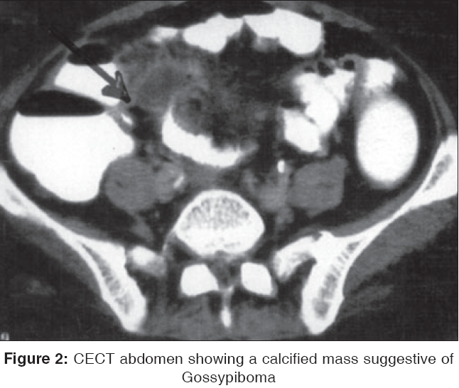
Indian Journal of Surgery, Vol. 68, No. 1, January-February, 2006, pp. 44-45 Images in Surgery Calcified abdominal mass Gogia Atul, Kakar A, Byotra SP Rajouri Garden, New Delhi Code Number: is06011 Case History A 35-year-old lady presented with pain abdomen right lower quadrant along with symptoms suggestive of subacute intestinal obstruction. She gave a past history of lower segment caesarean section 3 years ago. On examination, the abdomen was mildly distended and she had mild tenderness in the right lower quadrant. An X-ray of the Abdomen showed a calcified mass in the right lower quadrant [Figure - 1]. A contrast enhanced CT scan delineated the calcified mass in the lower abdominal cavity and was suggestive of a gossypiboma [Figure - 2] probably left during her previous surgery. She was operated and the sponge removed a repeat X-ray thereafter showed the clearance of the abdominal mass and the patient was relieved of her symptoms.Discussion Gossypiboma (from Latin gossipium cotton and Kiswahili boma place of concealment) or retained surgical sponge is a ubiquitous medical error that is avoidable. It can cause serious morbidity and possibly even mortality.[1] It should be considered in the differential diagnosis of acute mechanical obstruction in patients who have undergone laparotomy previously. It has been reported following surgical procedures such as abdominal, thoracic, cardiovascular, orthopedic, and even neurosurgical operations.[2] Although the real incidence is unknown, it has been reported as 1 in 100 to 3000 for all surgical interventions and 1 in 1000 for intra-abdominal operations.[3],[4] Non-specific clinical symptoms and inconclusive imaging findings may preclude an accurate diagnosis. The diagnosis is made easily in case there is suspicion and a plain abdominal X-ray may pick it up. USG is another diagnostic tool, which may well demonstrate foreign bodies. CT and MRI reveal comprehensive details about the mass in most cases. The best approach in the prevention of this condition can be achieved by meticulous count of surgical materials in addition to thorough exploration of surgical site at the conclusion of the operations and also by the routine use of surgical textile materials impregnated with radio-opaque marker. Surgery is the most reliable method for removing foreign bodies especially from the abdomen. Gossypiboma also carries some medico-legal implications; the presence of a foreign body inside the patient can be easily proved and they may litigate the responsible surgeon because this is an avoidable problem. Moreover, gossypiboma may be misdiagnosed as a malignant tumor and lead to unnecessary invasive diagnostic problem or extensive extirpative surgery, which may result in further complications.[5] References
Copyright 2006 - Indian Journal of Surgery The following images related to this document are available:Photo images[is06011f2.jpg] [is06011f1.jpg] |
| |||||||||
