 |
| About Bioline | All Journals | Testimonials | Membership | News |
|
||||||
|
||||||
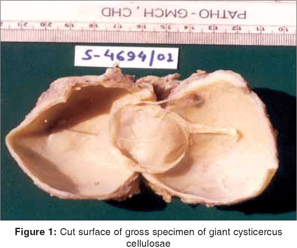
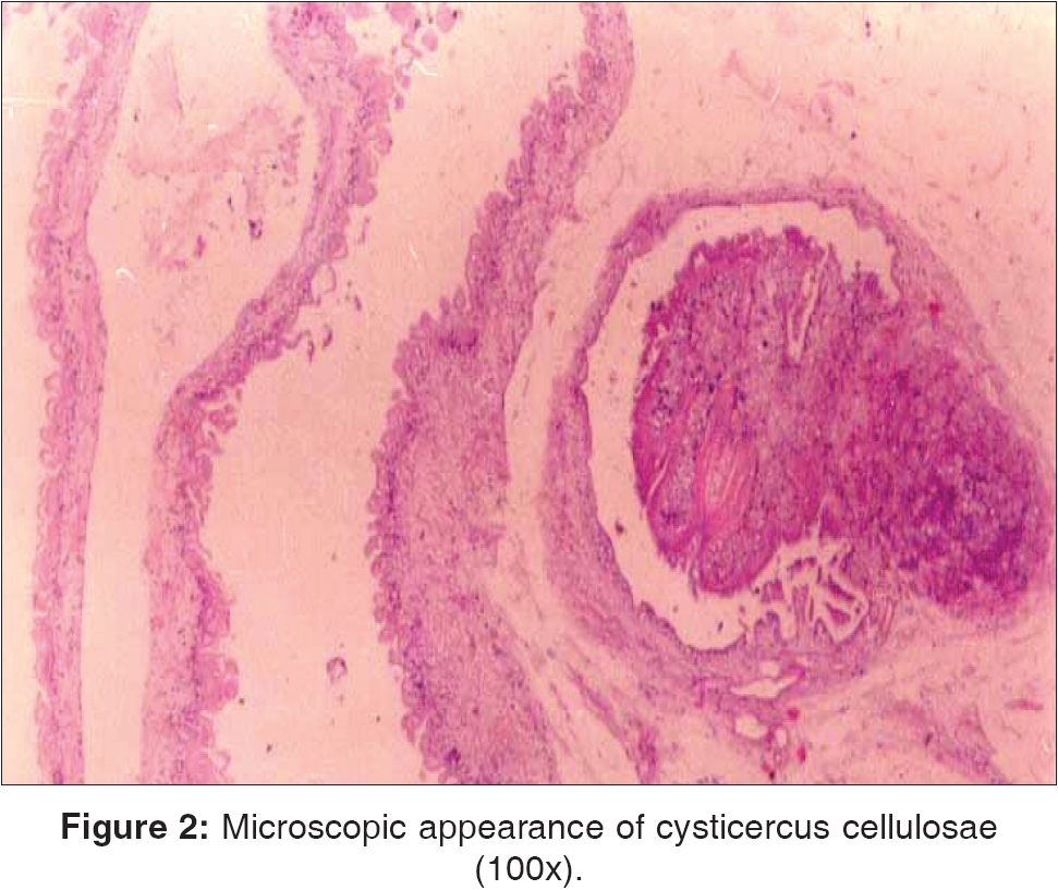
Indian Journal of Surgery, Vol. 68, No. 1, January-February, 2006, pp. 46-47 Images in surgical pathology Giant cysticercosis: An uncommon presentation of a common lesion Punia R. P. S., Mittal R, Amanjit, Mohan Harsh Department of Pathology, Govt. Medical College and Hospital, Sector-32 A, Chandigarh - 160030 Code Number: is06012 Human Cysticercosis, an infection caused by larvae of Taenia solium , is a public health problem in many developing countries. The larval stage of T. solium i.e. cysticercus is maximum up to 1 cm in size. Cysticercosis with giant cysts, which is relatively uncommon, has generally been defined by the presence of a cyst of more than 50 mm in diameter and 60 ml in volume.[1] Large cysticerci produce swelling mimicking tumours. We report a case of a 32-year-old female patient who presented with swelling in the right forearm for 5-6 months, which gradually increased in size. There was a history of pain for 1-2 months. On examination the swelling was 7x4 cm, fluctuant, cystic, mobile. The overlying skin was mobile, and there was no change of temperature nor tenderness. The swelling was not adherent to deep tissues. Intra-operatively, cystic swelling above the flexor muscles of the forearm was found. There was a clinical impression of hydatid cyst or benign soft tissue tumour. The cyst was 9 x 5.5 x 2 cm in size [Figure - 1]. On section clear fluid and dirty grey white semi-solid material came out. The lumen of the cyst contained a thin papery membrane. The wall thickness of the cyst was 0.2-0.3 cm. Microscopic examination revealed cysticercus cellulosae [Figure - 2]. The wall of the cyst showed palisading histiocytes and mixed inflammatory cell infiltrate. Giant cysticerci are usually larvocysts of Taenia hydatigenia and range in size from 0.8 to 8 cm.[2] Species identification on histologic examination is difficult especially in the absence of scolex and hooklets. Clinicians usually suspect hydatid cyst, haematoma or tumour in such a large cutaneous swelling. However, microscopic examination of the cyst can easily differentiate giant cysticerci from the laminated membrane of the hydatid cyst and can also rule out the clinical possibility of any tumour. References
Copyright 2006 - Indian Journal of Surgery The following images related to this document are available:Photo images[is06012f2.jpg] [is06012f1.jpg] |
| |||||||||
