 |
| About Bioline | All Journals | Testimonials | Membership | News |
|
||||||
|
||||||
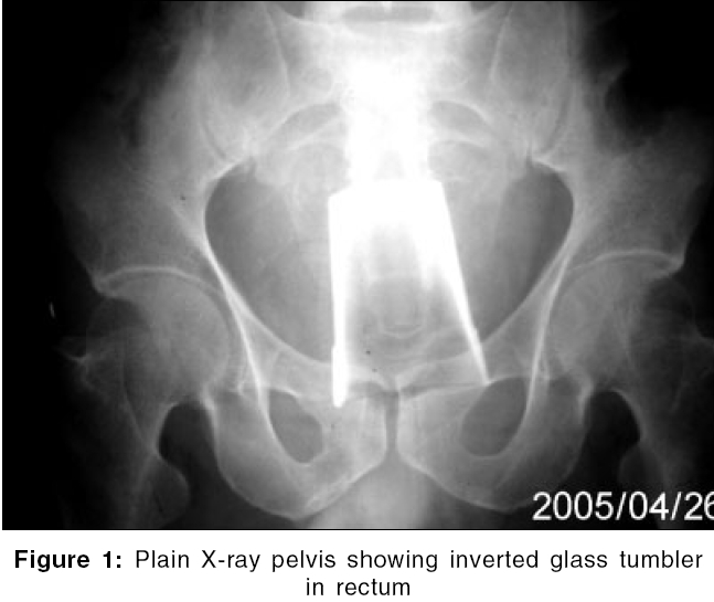
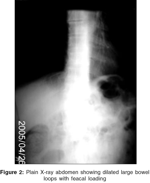
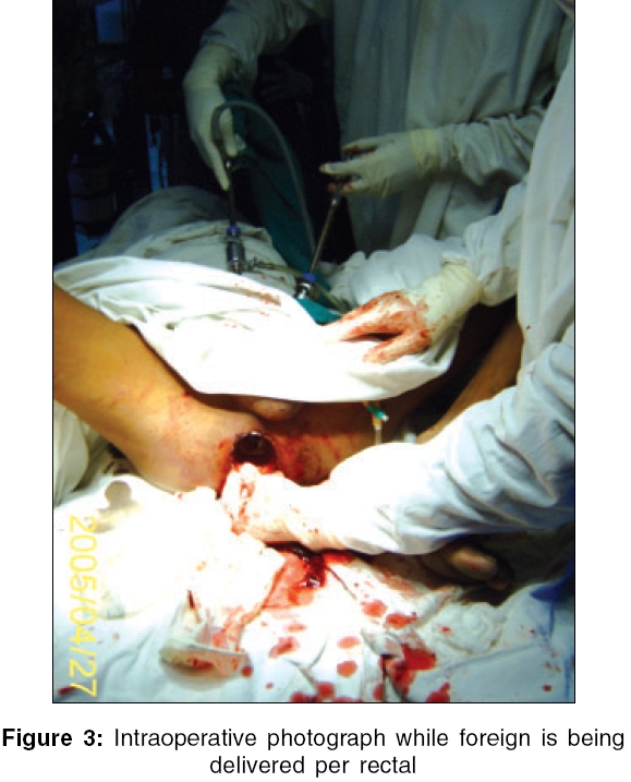
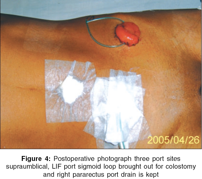
Indian Journal of Surgery, Vol. 68, No. 4, July-August, 2006, pp. 216-218 Case Report Laparoscopic assisted removal of rectal foreign body Bhanot Ashish, Patel GR, Bachani Mitesh, Gohil VijayrajD Department of Surgery, Govt. Medical College, Bhavnagar, Gujarat Correspondence Address:Ashish Bhanot, C-7 Doctors Quarters, Sir T. Hospital Campus, Bhavnagar - 364 001, Gujarat, India. E-mail: bhanotashish@rediffmail.com Code Number: is06061 Abstract 'Foreign' means originating elsewhere or simply 'outside the body.' Foreign body rectum is not as common as other parts of the body. Rectal foreign bodies present are difficult to manage. Emergency-department procedures include rectal examination, proctoscopy and abdominal radiography. Soft or low-lying objects having an edge could be grasped and removed safely in the emergency department, but grasping hard objects is potentially traumatic and occasionally results in upward migration toward the sigmoid. Although foreign bodies can be removed in the emergency department in about two out of three cases, some 10% still require a laparotomy and a diverting colostomy to remove the object or to treat bowel perforation. We are presenting a case of laparoscopic assisted removal of tumbler using 10 mm suction cannula to push the object down. Laparoscopy helped not only in retrieval but also enabled visualizing any bowel perforation due to foreign body and its manipulation.Keywords: 10 mm suction cannula, foreign body rectum, laparoscopic assisted, sigmoid colostomy Introduction Rectal foreign body, although infrequent, presents a challenge in management. Here is case, first of its type, where laparoscopy was used to assist retrieval of foreign body in the rectum as well as rule out any bowel perforation.Case report A 46-year-old male presented to the emergency department with complaints of bleeding per rectum. History of sexual assault by three truck drivers was there. When the patient refused homosexual relation, they forcibly introduced a glass tumbler in the patient′s rectum. Attempts were made to remove it by the local physician during which the margin of glass broke off and bleeding started per rectum. Per abdomen was soft and the patient also had left tunica vaginalis hydrocele. On rectal examination, drops of fresh blood were trickling from the anus. On finger examination, there was a reduced tone of anal sphincter and circumference of glass tumbler could be reached by the fingertip with difficulty. Proctoscopy confirmed presence of tumbler in the rectum, as open-end margins could be seen along with breach in anal canal, but the exact length could not be assessed. The patient was taken to the X-ray department and abdominal radiography was done. Radiograph confirmed the presence of radiopaque foreign body in the pelvis [Figure - 1]. There was no free gas under the domes of diaphragm [Figure - 2]. Abdominal ultrasound ruled out the presence of any free fluid in peritoneum. The patient was taken to the operative room with proper consent, including consent for colostomy. General anesthesia was given to the patient after the lithotomy position dilatation of anal sphincter was performed and the attempt for per rectal retrieval was tried but failed. Bimanually pushing object with abdominal pressure applied to facilitate caudal movement was tried but failed. Laparoscopy was planned on table but the patient had previous lower abdomen scar, so introduction of the first trocar was done by open method in infraumblical region. Other trocars were planned - one just on the right side of a point midway between umbilicus and pubic symphsis. Adhesions of previous surgery were there, which were dealt by blunt and sharp dissection. Another 10 mm port was planned at a point 3 cm medial to the left anterior superior iliac spine, keeping in mind the possibility of using it for sigmoid colostomy if required. Peritoneal cavity, sigmoid colon and rectum were inspected for any evidence of perforation by foreign body. Now the foreign body was located by indirect palpation using blunt suction tip. The tumbler was located and pushed down using constant pressure by blunt suction tip. At that time, fingers could be passed 1 inch beyond the margins of tumbler and the object was retrieved with great difficulty using fingers of both the hands [Figure - 3]. Rectum was palpated and inspected to rule out any breach. There was deep longitudinal tear in the lower part of the rectum, which required rest to heal, so diverting sigmoid colostomy was done. Sigmoid colon loop was brought out from port site in left iliac fossa and Ryle′s tube was kept as drain from 5 mm port site [Figure - 4].Discussion The objects homosexuals insert into their rectum are only limited by the capacity of their rectum, not their imagination.[1] Eighty percent of these events occur for sexual stimulation and in 10% cases, it is forcibly introduced during sexual assault.[2] These patients typically present to the emergency department in a delayed fashion because of embarrassment and often after multiple attempts at self-removal. A multi-disciplinary approach should be used when encountering patients with colorectal foreign bodies. Length of time since insertion and presence of rectal or abdominal pain, fever or rectal bleeding are important elements of the history. The keys to adequate care for these patients are respect for their privacy, determination of the type and location of the foreign body and determination if removal can be performed in the emergency department. Operating room procedures include anal dilatation under general anesthesia, transrectal manipulation, bimanual palpation if necessary and withdrawal of the foreign body.[3] The possibility of a perforation must be taken into account, especially with longstanding foreign bodies that can erode the bowel wall. A fewfree perforations are clinically obvious with free air on abdominal radiographs; but in few cases, small perforations sealed by omentum were not evident on radiographs. If there is difficulty in interpreting radiographs, an opinion from a radiologist should be obtained. Before attempting any manipulation in the operation theater, consent for laparotomy and colostomy should be sought. These patients are often deeply embarrassed and psychological support and confidentiality are essential. The role of the nursing staff involved in the care of the patient in such cases is highly important. The patient should be treated with bed rest, analgesia and mild sedation.[4] Soft- or low-lying objects having an edge could be grasped and removed safely in the emergency department, but grasping hard objects is potentially traumatic and occasionally results in upward migration toward the sigmoid. Operating room procedures include anal dilatation under general anesthesia, transrectal manipulation, bimanual palpation if necessary and withdrawal of the foreign body.[5] Frequently, delay in presentation and multiple attempts at self-removal lead to mucosal edema and muscular spasms, further hindering removal. Few cases require laparotomy, manually pushing object caudally towards anus. To avoid formal laparotomy and its morbidity, laparoscopy can be used as modality to push low and mid-high foreign bodies in rectum. Laparoscopy also helps to identify any sealed-off rectal perforation and the one which occurs during manipulation. Like any other minimally invasive procedures, it is patient-friendly due to small incision and early ambulation. The patient can be sent home early, thereby giving more confidentiality and lesser psychosocial trauma. In the era of minimally invasive surgery, this is another procedure that can be done by laparoscopy. References
Copyright 2006 - Indian Journal of Surgery The following images related to this document are available:Photo images[is06061f4.jpg] [is06061f2.jpg] [is06061f1.jpg] [is06061f3.jpg] |
| |||||||||
