 |
| About Bioline | All Journals | Testimonials | Membership | News |
|
||||||
|
||||||
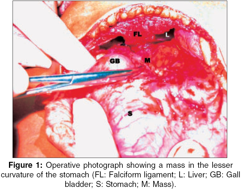
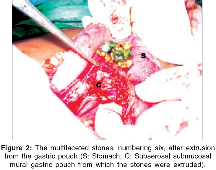
Indian Journal of Surgery, Vol. 68, No. 4, July-August, 2006, pp. 224 Images in Surgery Unusual presentation of Bouveret's syndrome Sarda AnilK, Bhalla ShwetaA, Lal Pawan, Neogi Sushanto Department of Surgery, Maulana Azad Medical College and Lok Nayak Hospital, New Delhi - 110 002 Correspondence Address:A. K. Sarda, 27 RPS, Triveni-1, New Delhi - 110 017, India. E-mail: aksarda@rediffmail.com Code Number: is06064 A 26-year-old female presented with symptomatic gallstones. An ultrasound examination showed multiple calculi in the gall bladder and a normal common bile duct. During elective cholecystectomy dense adhesions were seen around the fundus of the gall bladder in relation to the duodenum and distal stomach. The gall bladder was easily separated from the first part of the duodenum and the stomach. A black-coloured hard mass was noticed in relation to the lesser curvature and the gastrohepatic ligament, with no irregularity felt in the gastric mucosa. An incision was made along the mass for taking a biopsy. However, the hard mass was actually a collection of gallstones without any obvious communication with the gall bladder, completely covered by the gastric wall muscle and without any breach of the gastric mucosa [Figure - 1][Figure - 2]. The stones were removed and the gastric muscle closed in layers. Cholecystoduodenal and cholecysto-gastric fistulas causing gastric outlet obstruction (Bouveret′s syndrome) caused by gallstones or the classical gallstone ileus are known.[1] Entrapment of stones in the stomach is known to occur in up to 14% of all cases of gallstone ileus.[2] However, a review of the literature did not reveal any case report where gallstones were found in an intramural pouch of the stomach without any breach of the gastric mucosa and without any communication with the gall bladder. References
Copyright 2006 - Indian Journal of Surgery The following images related to this document are available:Photo images[is06064f2.jpg] [is06064f1.jpg] |
| |||||||||
