 |
| About Bioline | All Journals | Testimonials | Membership | News |
|
||||||
|
||||||
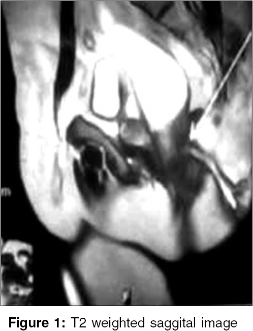
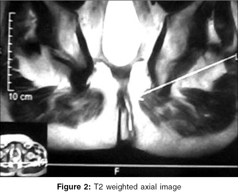
Indian Journal of Surgery, Vol. 68, No. 4, July-August, 2006, pp. 225 Images in Surgery MRI in fistula in ano K PraveenKumar, Satish Rao BS, Aithala SathyamoorthyP, Hanumanthappa MB, Vijaya G Department of General Surgery, Fr. Muller Medical College Hospital, Kankanady, Mangalore - 575 002, Karnataka Correspondence Address:Praveen Kumar K., Department of General Surgery, Fr. Muller Medical College Hospital Kankanady, Mangalore - 575 002, India. E-mail: drpkkumar@yahoo.com Code Number: is06065 A 57-year-old male patient presented with discharge from an opening on the left side of anus since 2 months. Examination revealed a fistula in ano, with the external opening 3 cm from the anal verge on the left posterolateral aspect and the internal opening was above the dentate line posteriorly. A fistulogram was performed, which suggested a high anal fistula. Magnetic resonance imaging (MRI) of the fistula in ano was done and this revealed it to be a low anal fistula [Figure - 1][Figure - 2]. The patient underwent a simple fistulectomy and recovered well. Fistula in ano is a commonly encountered problem in surgical practice. It is difficult to determine the exact anatomy of fistulous track by clinical examination alone. Fistulography, which has been used widely for distinguishing between high and low fistulas, has its own disadvantages.[1] Hydrogen peroxide enhanced 3-D endoanal ultrasonography and MRI are now being evaluated as the investigative modality. T2 weighted spectral fat saturation inversion recovery (SPIR) sequences and the conventional short tau inversion recovery (STIR) sequences of MRI have been described. While both these techniques are adequate to classify fistula in ano, it is easier with STIR due to superior resolution of pelvic floor structures.[2] T1 weighted sequences are generally noncontributory. References
Copyright 2006 - Indian Journal of Surgery The following images related to this document are available:Photo images[is06065f2.jpg] [is06065f1.jpg] |
| |||||||||
