 |
| About Bioline | All Journals | Testimonials | Membership | News |
|
||||||
|
||||||
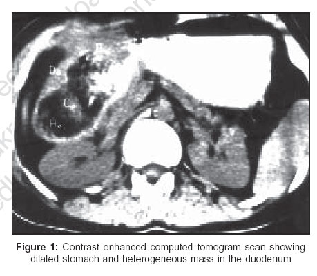
Indian Journal of Surgery, Vol. 68, No. 5, September-October, 2006, pp. 279 Images Gossypiboma eroding into the duodenum Rajeev Sharma, Atul Sachdev*, Sanjay Gupta, Robin Kaushik, Ashok Attri Departments of Surgery and *Medicine, Government Medical College and Hospital, Chandigarh, India
Code Number: is06080 The problem of retained surgical sponges (gossypiboma) in the abdominal cavity is not only a cause of morbidity in clinical practice, but also assumes great medicolegal significance in today′s era. An unusual presentation of gossypiboma is reported and the condition is briefly reviewed. A 26 - year-old female presented with features of gastric outlet obstruction after two months of open cholecystectomy. Upper gastrointestinal endoscopy revealed a grossly dilated stomach, with an eccentric pylorus that could not be negotiated. Contrast enhanced computerized tomogram scan revealed dilatation of the stomach and a mass in the first part of the duodenum, with very little oral contrast going beyond it [Figure - 1]. Exploratory laparotomy was planned. Peroperatively, the wall of the distal stomach and the duodenum was seen bulging outwards. A gastrostomy was done and a surgical sponge was visualized that was removed from within the duodenum through the pylorus. The patient did well in the postoperative period and was discharged on the eighth day after the surgery. Gossypibomas are surgical sponges that are retained inadvertently after surgery. Although they commonly remain asymptomatic, they may, at times present with variable symptomatology depending upon their site and the extent of inflammatory reaction. Thus, they may present as intra- or retro-peritoneal ′tumors′, abscesses, fistulae, extrusion through laparotomy wounds and uncommonly, as intestinal obstruction after intraluminal erosion into a segment of adjoining bowel.[1] The various radiological features of retained surgical sponges may include [2],[3] visualization of a radio opaque marker on plain X-ray, a well-defined hypoechoic mass with posterior acoustic shadowing or, a cystic mass with irregular internal echoes on ultrasound. The characteristic appearance on a CT scan is that of a well-defined heterogeneous mass with air bubbles, calcification and a wavy striped and / or spotted appearance or a whorled pattern. In this case, the sponge probably eroded into the duodenum slowly over a period of time, leading to a gastric outlet obstruction. It was only during surgery that the correct diagnosis could be established. References
Copyright 2006 - Indian Journal of Surgery The following images related to this document are available:Photo images[is06080f1.jpg] |
| |||||||||
