 |
| About Bioline | All Journals | Testimonials | Membership | News |
|
||||||
|
||||||
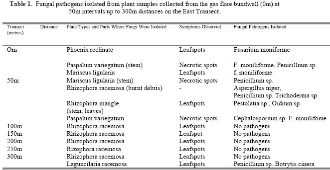
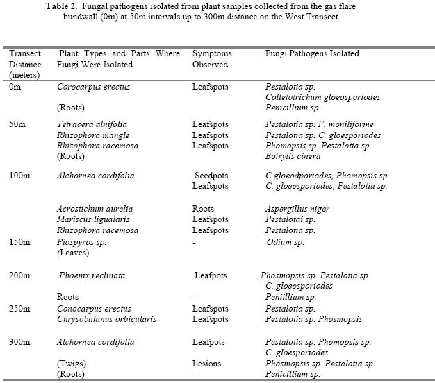
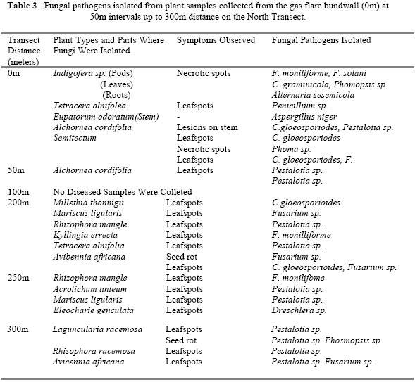
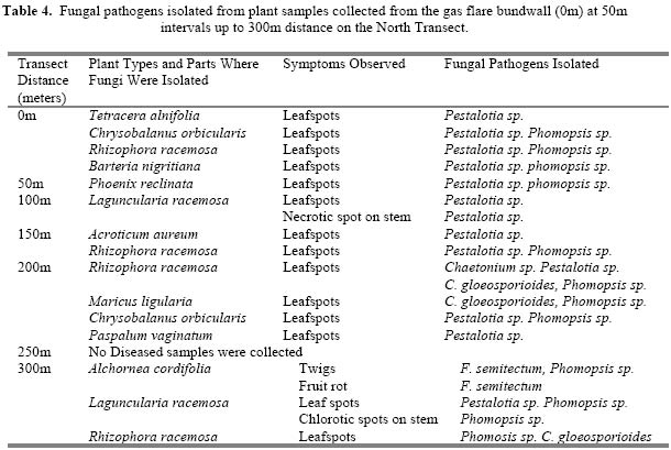
Journal of Applied Sciences & Environmental Management, Vol. 9, No. 1, 2005, pp.69-73 Health Impact Assessment of Mangrove Vegetation in an Oil Spilled Site at the Bodo West Field in Rivers State, Nigeria UMECHURUBA, C.I Environmental Impact Assessment Unit, Department of Health, Safety and Environment, Shell Petroleum Development Company Ltd., P.O. Box 263, Port Harcourt, Nigeria. Code Number: ja05012 ABSTRACT: A post-impact health assessment of the mangrove vegetation of Bodo West Field in Rivers State of Nigeria where three oil spillages occurred was carried out to determine any major changes from the baseline profile of the area 10yrs after spillage. The accidents resulted in the discharge of a total of 1860 barrels of crude oil, which cut fire as it spilled destroying the mangrove vegetation. Diseased plant samples were collected from infected plants at 50m intervals to a distance of 300m from the point of spillage along four transects in the East, West, North and South directions. Results of laboratory diagnosis showed that most of the plants especially plants along the South and East transects were heavily infected with necrotic leafspots caused mainly by Pesalotia and Phomopsis spp. Rhizophora mangle, Rhizophora racemosa and Avicennia africana were the most heavily infected plants. Other genera of fungal pathogens isolated were Alternaria, Aspergillus, Botrytis, Chaetonium, Colletotrichum, Dreschlera, Fusarium Macrophomina, Penicillium, Phoma and Trichoderma. No plant pathogenic bacteria or viruses were isolated. Oil pollution predisposed the plant to fungal disease attack and also impacted the soil and vegetation. @JASEM
This study was therefore undertaken to determine the impact of oil pollution on the health of mangrove vegetation in the Bodo West in the tidal plain of the Bonny River in River State which has suffered three major oil spills. MATERIALS AND METHODS Field Sampling: Four transects were cut from the edge of the gas flare bundwall of Bodo West Flow Station in the East, West, North and South directions up to a distance of 300m from the oil spilled zone. Plant samples (roots, stems, leaves and seeds) of different plant genera and species were taken on a daily basis along the transect lines for laboratory diagnostic studies. Five diseased plants of each plant type seen in the surrounding area of 0m, 50m, 100m, 150m, 200m, 250m, and 300m distances of the four transects were collected. Samples collected at the 300m points served as controls since the oil spread did not reach there. Field sampling took 7days to complete. Plant samples were taken to the laboratory and kept in the refrigerator until when used. Laboratory Studies: For fungal diagnostic studies, acidified potato dextrose agar (APDA) medium was used initially but was later discontinued due to lots of contamination by saprophytic fungi. The Standard Blotter Method (ISTA, 1999) was used instead. The leaves of each plant type were cut to pieces of 0.5 to 1.0cm and the stems and roots were cut to pieces of 1.0 to 1.5cm making sure that the healthy and diseased areas were included in each piece. The seeds were not excised. Tissues pieces of the same plant type and part were pre-treated with 1% sodium hypochlorite for 3minutes. Tissues were rinsed in sterile distilled water before plating each piece on three layers of moist sterile filter papers in each Petri dish. Five pieces were plated per Petri dish, making sure that the pieces were well separated. Five Petri dishes were used for each plant part. The tissue pieces were then incubated in an incubator a 22°C ± 2°C for 7days for fungal organisms to sporulate. Tissues were examined under a stereobinocular microscope (5-50X magnification) for spores and fruiting bodies for identification of fungi. Temporary spore mounts of each observed fungus were also made and observed under a light compound microscope (5-100 x-magnification) to confirm identification to genus and species level. Fungal pathogens identified according to plant type and parts were recorded. For bacterial studies, dipping them in 70% ethanol disinfected several pieces of younger infected leaves of each plant type and rinsing three times in sterile distilled water. The pieces of leaves were put in a sterile blender and blended for 2minutes to release the sap. One millilitre of the sap solution (stock solution) was used in dilution series of 10-1 to 10-7 in test tubes containing 9ml of sterile distilled water. One millilitre of 10-6 and 10-7 dilutions were plated onto nutrient agar (NA) medium in Petri dishes. Five Petri dishes were used per plant part. With a sterile L-shaped glass rod, the solution on the medium was evenly distributed. Dishes were inverted after the solution had settled on the agar and then incubated at 30°C ± 2°C for 1-3days for bacteria growth. Dishes were examined each day and observations recorded. In case of isolation of seed-borne pathogenic bacteria, 50g of seed types were soaked in 120ml of sterile distilled water for 24hrs at 5°C (refrigerator). From soaked seeds solution, a dilution series of up to 10-7 was made as previously described. One millilitre of each seed-type solution (10-6 or 10-7) was spread onto the NA medium in Petri dishes. Five Petri dishes were used for 10-6 or 10-7 dilution per seed type. After allowing the solution to settle, the dishes were incubated at 30°C ± 2°C for 1-3days. Dishes were examined each day for bacteria growth and observations recorded. RESULTSFungal diseases recorded among the various plants sampled along the 4 transects in the oil-polluted area of Bodo West Field are shown in Tables 1,2,3 and 4. The main fungal genera commonly associated with the various plant types and parts sampled include: Aspergillus, Cephalosporium, Colletotrichum, Fusarium, Penicillium, Pestalotia, and Phompsis. Disease incidence and severity were greatest in the East and South transects that were heavily polluted with crude oil. A vast area of the mangrove vegetation from the gas bundwall up to 200m sites of the East and South transects was burnt by the heat emanating from the flare. Symptoms observed on surviving diseased plants in the area include: necrotic leafspots, chlorosis, premature abscission and dieback. Virtually all the plants in the two transects had several necrotic leafspots caused by Pestalotia sp. and Phomosis sp. Rhizophora mangle, Rhizophora racemosa and Avicennia africana were the most severely infected plants. In addition, the herbicidal effect of the crude oil (Hutchinson and Freedman 1978) contributed to the disease severity. Fungal disease incidence and severity on plants at 300m distance from gas flare bundwall in all the four transects were very low. These areas served as controls. DISCUSSION Campbell et al. (1972) isolated Cephalosporium spp. from oil-polluted soil and Odu et al. (1985) isolated Aspergillus, Cladosporium, Fusarium, Penicillium, Trichoderma and Rhizopus in oil-polluted soil. These findings are in agreement with the findings of this study. These genera of fungi seem to have the ability to utilise crude oil hydrocarbons as source of carbon and energy for growth. The crude oil does not seem to affect their pathogenic potential. Okafor (1987) reported that crude oil did not have any effect on the pathogenic potential of seed-borne fungi such as Fusaium moniliforme and Cephalosporium acremonium in maize seeds soaked in weathered crude oil for 3 months. Phytotoxicological effects of the crude oil on the structure and physiology of the plants seemed to have contributed immensely in predisposing the plants to the pathogens. Host resistance to infection was weakened and disease development enhanced. Crude oil in oil-spilled area usually penetrates the soil and directly affects the microbial activities, dissolved oxygen level and plant root system (Petts and Eduljee, 1994). It causes the soil particles to stick together thereby decreasing the porosity of the soil. It increases the acidity of the soil thereby affecting the rate of mineral nutrients uptake by plants (Petts and Eduljee, 1994). It also affects the population of some beneficial microorganisms in the soil. Soil acidity caused by oil pollution is known to inhibit non-symbiotic nitrogen fixation by Azotobacter spp. as well as symbiotic nitrogen fixation by Rhiobium spp. (Munns, 1965). All the above-mentioned factors may have contributed directly or indirectly to the aberrant metabolism of the plants thereby reducing the resistance of the plants to attack by plant pathogens. In addition, the herbicidal effects of the crude oil may have contributed to the disease severity (Hutchinson and Freedman, 1978). The soil and vegetation of the area were heavily impacted by the spills. There is need to re-vegetate the affected area to restore the field to it's former glory. REFERENCES
The following images related to this document are available:Photo images[ja05012t2.jpg] [ja05012t3.jpg] [ja05012t1.jpg] [ja05012t4.jpg] |
| |||||||||
