 |
| About Bioline | All Journals | Testimonials | Membership | News |
|
||||||
|
||||||
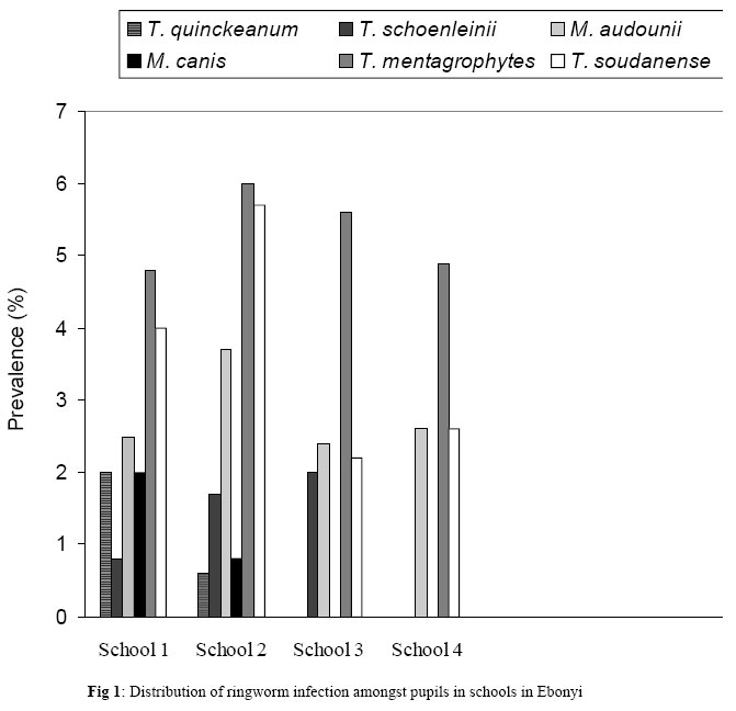
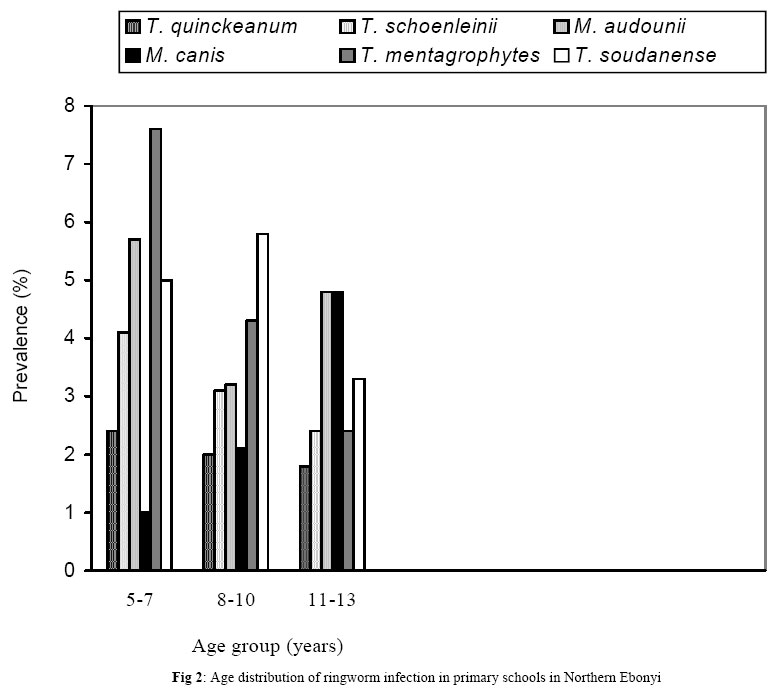
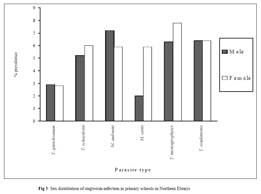
Journal of Applied Sciences & Environmental Management, Vol. 9, No. 3, 2005, pp. 21-25 Prevalence and distribution of ringworm infections in Primary School Children in parts of Eastern, Nigeria *1ANOSIKE, J C; 1KEKE, I R; 2UWAEZUOKE, J C; 2ANOZIE, J C; 2OBIUKWU C E; 2NWOKE, B E B; 2AMAJUOYI, O U 1Department of Animal and Environmental Biology, 2Department of Microbiology ImoStateUniversity, Owerri.p. M.B. 2000 Owerri, Nigeria E-mail: jc_anosike@yahoo.com Tel No. – 2348037235456 *Corresponding author: E-mail: jc_anosike@yahoo.com . Tel No. – 2348037235456 Code Number: ja05053 ABSTRACT: A study on the prevalence and distribution of ringworm infection amongst primary school children in northern EbonyiState of eastern Nigeria was carried out between November 2002 and June 2003. Of the 279 pupils sampled randomly from four schools, 59 (21.1%) had ringworm infection. While only two genera of fungi Microsporum and Trichophyton were isolated, six species viz: M. canis (11.9%), M. andoninii (20.3%), T. sondamense (20.3%), T. mentagrophytes (22.0%), T. schoenleinii (15.3%) and T. quinckeannan (10.2%) were also encountered. The distribution of ringworm among schools was not statistically significant (P > 0.05). Both male and female pupils with in the age bracket of 5-7 years were significantly infected than other age categories (P<0.05). Infection decreased with increase in age. Sex-related prevalence was not statistically significant (P>0.05). M. andoninii was more predominant over others in males while T. mentagrophytes was more prevalent in female pupils. Poor infrastructures (houses and classroom), children playing with animals, the Nigerian environmental vis-a-vis personal uncleaniness by pupils are contributing factors to the high frequency and severity of ringworm in the area. @JASEM Ringworm infection medically known as dermatophytosis caused by dermatophytes which are highly specialized group of fungi. They affect the superficial keratinzed tissue (skin, hair and nails) of man and animals. It is a common superficial fungal infection found throughout the world (Kern 1985). It occurs primarily in prepubatal children over the age of 6 months (Ekeski and Hay; 1996). It is highly contaginous and represents a significant public health problem, particularly among school children (Fatini and Al-Samarai 2000; Omer 2000; Higgins et al 2000). Ringworm infection is not a reportable disease but is a cause for concern because of its contaginous nature. It can be transmitted though body contacts (person to person transmission) mainly in refuge carrup or schools or through inanimate objects like cloths combs or hair dressers equipment. Dermatophytosis can be unsightly or disfiguring causing varing symptoms depending on the part of the human skin they are found. For instance, ringworm of the skin (Tinea corporis) causes annular lesions with a clearing, scaly center surrounded by a red advancing border that may be dry or vesicular. Ringworm of the scalp, (Tinae capitis) also gives rise to dull gray, circular patches of alopecia, scaling, itching and black dot (Jawetz et al 1998). Though other ringworm species abound, the present work is restricted to Tinea capitis and T corporis. This disease presently is one of the most prevalent dermato mycoses in Nigeria and represents a major public health in children of school age. Published reports on this disease in Nigeria is scanty (Egere and Gugnani, 1980). The aim of the present study therefore was a mycological assessment of pupils in northern Ebonyi state Nigeria to estimate the prevalence and distribution of ringworm infection. MATERIALS AND METHODS The Study area: Four communities of Ebonyi Local Government area of Ebonyi State, Nigeria were chosen for this research and four mixed schools were randomly selected. These schools include: ItemCommunityPrimary School (school 1), NdufuCommunityPrimary School (school 2), UgboenyiCommunityPrimary School (school 3), and OjianyaCommunityPrimary School (school 4). Ebonyi Local Government area is situated in the North Eastern part of EbonyiState. It also shares boundaries with Benue and CrossRiverStates as well as Izzi and Abakiliki Local Government areas respectively. EbonyiState occupies the area lying between co-ordinates 6° 15’ and 5° 36’N and between 7° 30’ and 8°’ 18’E. Study Population: The Local Government Area is made of several inhabitants who are mainly farmers. The people depend mainly on pond water for their domestic uses and these ponds as were noticed, normally get dry during the dry season. There are fewer blockhouses, most of the houses were of mud (that is, little huts). Most of these blockhouses including classrooms are poorly constructed, uncompleted and many are not even cemented, paving way for the children to be in constant contact with the soil. Also, most of the schools visited had no seats and the pupils were found sitting on the un-cemented ground copying lesson notes. Ringworm Survey in the Study Area: This research work lasted for six months. The need and importance of the study was also highlighted to the officers. The heads of the primary schools were informed on the work schedule. The pupils were given health education on the disease. All areas of each pupil’s head scalp, chest, hands, legs and other parts of the skin were thoroughly examined for evidence of scaling, crusting, follicular inflammation and hair loss of erythema. In each clinically diagnosed case of Tinea capitis and Tinea corporis, a detailed history was recorded. Information was noted on the disease duration, socioeconomic status and the level of crowding at home. Pupils were asked about their bathing habits (the number of times they bath in a day, use of soaps and sharing of towels with their relation or friends). They were also asked about their habits of sharing clothes, caps or hats at home and in school and the existence of potentially contagious contacts, including contact with animals. Family history and personal hygiene were also taken into consideration. Sample Collection and Methods: In all suspected cases of T. capitis and T. corporis, the diseased areas of the skin or head were thoroughly cleaned with alcohol and hairs and scales were collected for mycological examination using the most appropriate technique used by Fathi et al (2000). Hairs from the diseased areas were examined with the aid of woods’ light for positive examinations, which were confirmed with the presence of green fluorescence (Ogbonna et al 1986). Such fluorescing hairs were then taken. Where negative results were obtained with wood’s light, dull gray hairs were collected. The pieces of hair were also collected with sterilized containers. The scrapings were handled separately and no individual scraping was allowed to mix up with the other. The scrapings and the pieces of hair were plated out separately on sabourand agar, and cycloheximide media (Mycosel or Dermatophyte test medium). Cycloheximide was employed because saprophytic fungi and yeasts normally present as contaminants were inhibited by cycloheximide. Also media with antibacterial antibiotics greatly facilitate the isolation of fungi from non-sterile specimens. Four to five small pieces of hairs collected from the diseased areas of a particular individual were distributed evenly over the surface of a plate of agar. The resultant culture plates were incubated at 27°C for 4 weeks and then examined for the presence of dermatophytes. Subculture was made on SDA for further identification after the growth of the dermatophytes was established. Both net and slide culture techniques were carried out as described by Omar (2000). RESULTS Of the 279 pupils examined for ringworm infection, 144 and 135 were males and females respectively. Altogether, 59 (21.2%) were infected. Two genera (Microsporum and Trichophyton) and six species were isolated (Table 1). T. mentagraphytes was the highest in terms of prevalence and distribution, followed by T. sondanense, M. andounii, T. schoenleinii, M. canis with T. quinckeanum as the least. Figure 1 depicts the distribution of ringworm infection amongst pupils Ebonyi schools. Prevalence and distribution of the disease varied among various schools. T. mentagraphytes had the highest. Overall age prevalence and distribution of ringworm infection among pupils in primary schools in northern Ebonyi showed that infection decreased with increase in age. Infection was higher in the 5-7 year age group, followed by 8-10 years age bracket with those beyond this age group as the least (Figure 2). Sex distribution of ringworm infection among pupils in primary schools in northern Ebonyi showed that although more females than males were infected, it was not statistically significant (P>0.05). Details are shown in Figure 3. Table 1: Gender and age-related distribution and prevalence of ringworm infection in NorthernEbonyiState
DISCUSSION Ringworm is a common dermatophyte infection that constitutes an important public health problem among children worldwide, including Nigeria (Ive, 1966; Egere and Gugmani 1980; Ajao and Akintude 1985; Ogbonna et al., 1986). The disease remains endemic in Nigeria, largely because of lack of information on its prevalence and the absence of control measures. The present study revealed that 21.1% of primary school children in northern Ebonyi of Ebonyi state, Nigeria were infected by species of superficial dermatophytes. This finding agrees with other reports in Nigeria. However, much attention has not been given to ringworm infections. Of the dermatophytes isolated, M. andoninii with a prevalence of 20.3% is an anthropophilic species. It occurs mainly in prepubatal children. T. soudanense which was recovered from the schools children (20.3%) is an endothrix species and believed to be endemic in central and West Africa (Ogbonna et al., 1986). M. canis with a prevalence of 11.9% in the present study must be have originated from the bodies of infected animals. This fungus is believed to be zoophilic and plausibly originated from nature cats, dogs, cow and other animals since people in the area share their residential houses with domestic animals. Both T. mentagraphytes and T. schoenleinii are also anthropophilic species and the former is the most prevalent and it is of great public health importance. One of the greatest problems hindering the prevention and eradication of ringworm infection is the presence of healthy asymptomatic dermatophyte carriers. Majority of the pupils examined herein showed no physical symptoms of infection, yet samples collected from some of these asymptomatic pupils yielded significant growth of dermatophytes. This observation is in line with the reports of Ive (1966) who found that asymptomatic carriers of dermatophytes may be equal to symptomatic sufferers. This should keep both parents and teachers at alert so that adequate preventive measures would be taken to reduce the rate at which infection spreads in the schools amongst pupils. The prevalence and distribution of ringworm infection observed herein (21.1%) is relatively high compared with either the report of Ajao et al (1985) amongst should children in Ile-Ife, Nigeria (14.02%) or those of Omar (2000) in Alexandria (7.4%) as well as Fatini and Al-Samarai (2000) in Iragi children (2.7%). The difference may be due to variation in environmental and climatic conditions of the areas studied. Gender-related studies on the prevalence of ringworm in Nigerian has been fragmentary (Ogbonna et al 1986). Here, more females than males were infected though this was not statistically significant similar to the reports of Omar (2000) in Alexandria. This finding suggests that the infection is related to personal hygiene and its prevalence can be reduced by adequate health education and good personal hygienic practices. Higher prevalence of infection was found amongst children under the age of 10 years than older ones, suggesting that ringworm is mainly a pre-pubatal disease. Other researchers, Figueroa (1997) in South Western Ethiopia, Venugopal and Venugopal (1993) in Saudi Arabia, Addel-Hafez (1997) in Sohag governorate as well as Omar (2000) in Alexandria have subscribed to this age differential. This can be explained by poorer hygiene at this age as well as the absence of saturated fatty acids that provide a natural protective mechanism (Fisher and Cook 1998). Poor infrastructures (houses and classrooms) are contributing factors to the high prevalence of dermatophytes amongst school children in northern Ebonyi. In the four rural schools sampled, they lack good accommodation for studying. They sat on the uncommented floors where side walls are made of palm leaves. Children contact the infection from the soil (Ogbonna et al 1985). Mercantini et al (1980) recorded high frequency of Karetinophilic fungi from floors in RomanPrimary Schools. The playing habits of these children always bring them in constant contact with the soil. The habit of accompanying their parents to the farm also brings these children in close contact with the soil. Most pupils examined seldom had regular bath and the fungal spores once deposited on the skin from the soil have ample chance of germinating and colonizing the skin. Children were being seen playing with animals such as cow, goat, sheep, cats and local dogs which are known sources of infection (Fathi et al 2000; Ogbonna et al 1986). Therefore, the Nigerian environment vis-a-vis personal uncleaniless are contributing factors to the high frequency and severity of ringworm infection in Ebonyi state. This calls for good leaning environment and intensive health education of the school children on personal cleaniless. In nature, man and animals could act as important reservoirs of skin diseases. The importance of human and animal presence in the environment in the sense of direct interaction between human and animal ‘pressure’ and the presence and the distribution of these fungi in the environment itself have been reported (Mercantini et al 1980). They observed that this interaction is in turn determinant of the epidemiology of diseases caused by Keratinophilic fungi. The phenomena determining the ecology and total numbers of fungi on the skin depends on the amount of moisture present. Apart from the importance of water, the relative importance of other factors in promoting or inhibiting fungal growth on skin will depend on the species or even the strain under consideration. The skin is an environment unfriendly to most microbial species but sufficient nutrients are available to allow a small number to survive and multiply (Ogbonna et al 1986). However, undesirable microbial invaders could be discouraged from colonizing the skin of animals by either the presence of the free fatty acids of the skin, surface lipids, low hydration of the stratum corneum or the presence of bacteriocius as well as other inhibitors produced by the resident microflora. This is true of dermatophytes causing ringworm infections. In conclusion, the present work has revealed the existence of ringworm (Tinea capitis and Tinea corporis) among the population at risk. The school children and their teachers were not aware of the existence of the disease, hence the infected children represent a persistent and hidden source of infection. Therefore, routine regular inspection of school children on hygiene is recommended. Adequate and quality health education should be mounted in schools in EbonyiState in particular and Nigerian Schools at large. Parents are adviced to ensure that their children maintain both personal and environmental hygiene at homes. Also, playing with animals by school children should be discouraged. Inaddition, government should provide good learning infrastructures at school especially rural schools where pupils resort to improvise which exposes them to risks of infection. Finally, combined effort of the school children, teaches, parents as well as the local, state and federal (National) governments in the promotion of health education in schools, personal and community hygienic practices and provision of good infrastructures both in schools and residential areas would provide a conducive environment free from ringworm infections. Acknowledgement: We acknowledge with thanks the assistance received from Prof. B.E.B Nwoke and Dr. I.N.S. Dozie of Imo State University Owerri during the field and Laboratory activities. Encouragements received from Mr and Mrs S.O. Anozie are appreciated. REFERENCES
Copyright 2005 - Journal of Applied Sciences & Environmental Management The following images related to this document are available:Photo images[ja05053f3.jpg] [ja05053f2.jpg] [ja05053f1.jpg] | |||||||||||||||||||||||||||||||||||||||||||||||
| |||||||||