 |
| About Bioline | All Journals | Testimonials | Membership | News |
|
||||||
|
||||||
Journal of Applied Sciences & Environmental Management, Vol. 9, No. 3, 2005, pp. 115-119 The antisickling effects of dried fish (tilapia) And dried prawn (Astacus red) 1NWAOGUIKPE, R N; 2*UWAKWE, A A 1Department
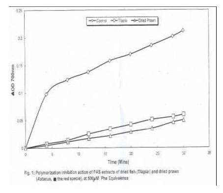
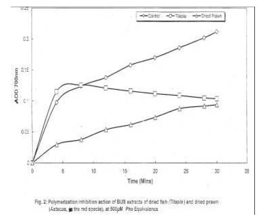
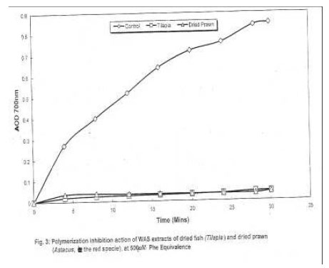
of Biochemistry, Federal University of Technology Owerri, Nigeria Code Number: ja05070 ABSTRACT: The antisickling effect of dried fish (Tilapia) and dried prawn (Astacus red) were investigated to ascertain the ability of the extracts of these samples to inhibit polymerisation of sickle cell haemoglobin (HbS), improve the Fe2+/Fe3+ ratio and lower the activity of lactate dehydrogenase (LDH) in blood plasma. The samples were first ground into powder and soaked in chloroform/dichloromethane to defat them and in essence produce the fat soluble fraction (filtrate). The defatted residues were soaked in methanol for 24 hours to obtain a methanol soluble fraction. This was finally fractioned in a mixture of BuOH/H2O (1:1) to give the butanol-soluble (BUS) and water-soluble (WAS) fractions respectively. These fractions were subsequently concentrated by rotary evaporation. The fat-soluble (FAS), BUS, and WAS phases were able to inhibit HbS polymerisation to varying degrees from 50% for FAS to 95% for BUS. The water-soluble phases of these samples were also found to increase the Fe2+/Fe3+ ratio from 6% to 95%. The phases equally reduced LDII activity in serum of ten sickle cell disease patients to varying degrees from 12% to 40%. Nutritionally, the different fractions or phases were found to be rich in free amino acids which ranged from 951.05mg/100g of sample for tilapia to 1906.05mg/100g of sample for crayfish (Astacus; red). The soluble protein concentration of the samples was equally estimated. Dried tilapia has an aggregate protein content of 28.7.30mg/100g of sample while dried prawn has 1626mg/100g of sample. Dried fish (Tilapia) and dried prawn (Astacus red) could both be nutritionally and therapeutically beneficial in the management of sickle cell disease. @JASEM Hemoglobin S differs from HbA in the substitution of valine for glutamic acid in the sixth position of the β-chain amino acid sequence. Hemoglobin HbSS, in whom all the hemoglobin type is HbS, always manifest features of sickle cell disease, which may be fatal in childhood. Sickling of red cells occur as a result of polymerization of deoxygenated HbS molecules, so that, they become stacked linearly. Clinical symptoms occur in homozygotes and develop at about 6 months old. There is a chronic hemolytic anemia and recurrent painful vasocclusive crises because of the sickled erythrocytes blocking small vessels. This leads to tissue ischaemia and infarction, mostly affecting the liver, spleen, lungs, brain and retina. Leg ulceration and priapism are also evident. These crises may be precipitated by minor infections, severe cold, exercise, dehydration and pregnancy. Several therapies have been prognosed and many chemical substances investigated for their possible role in the management of sickle cell disease. However this disease remains one chronic disease in which the role of nutrition in its aetiology has not been systematically addressed. Many investigations have been carried out on the role of some dietary supplements, such as thiocyanate (Agbai, 1986). Lactate dehydrogenase (LDH), is a sensitive indicator of hemolysis. Its level in sickle cell blood correlates with the severity of crises. Different preparations such as hydroxyurea, erythropoietin and tucaresol, have been found to reduce the level of serum LDH activity, and bilirubin, and increase the level of fetal hemoglobin, HbF. (Goldberg et al, 1992; Roopen, et al 1996). Different species of legumes abound in tropical Africa, which are very rich sources of proteins and amino acids. Because of the antisickling effects of certain amino acids such as phenylalanine, lysine, arginine etc, (Noguchi and Schetcher, 1977; Ekeke and Shode, 1990); we were prompted to investigate, the antisickling potency of some fish and crayfish samples, which abound in our lakes, rivers and oceans as rich sources of amino acids and proteins. MATERIAL AND METHODS Dichloromethane, chloroform, methanol, butanol, LDH kit, Ninhydrin, were all purchased from Sigma Biochemicals, London. The fish samples were purchased from a local market in Port Harcourt, Nigeria. Blood samples used were collected from ten sickle cell patients of ages 14 – 25 years and of both sexes (6 males and 4 females). The blood samples were confirmed as HbSS samples using haemoglobin electrophoresis. Portions (0.2cm3) of the whole blood samples were used for Fe2+/Fe3+ experiment while the remaining portions were collected into citrate anticoagulant tubes. Erythrocytes were isolated from the blood samples by centrifugation at 10,000g for fifteen minutes using bench centrifuge (MSE minor). Following careful siphoning of the plasma (with a Pasteur pipette), the erythrocytes were by repeated inversion, suspended in a volume of isotonic saline (0.90% NaCl) equivalent to the volume of the siphoned plasma. The erythrocyte suspension was then frozen at OoC and subsequently thawed out to produce a haemolysate for the haemoglobin polymerisation experiment. Extraction of Fat-Soluble (FAS) components: The amounts (200g) of the dried fish (Tilapia) and dried prawn (Astacus red) were separately ground with a grinder and blender and the resultant samples soaked in 400ml of chloroform/dichloromethane for twenty four (24) hours to defat them and in essence generate the fat-soluble fraction by filtration. The residue was kept for methanol extraction while the filtrate was subsequently evaporated using rotary evaporation and the resulting fat-soluble (FAS) extract weighed. Methanol Extraction Process: The residues from the chloroform/ dichloromethane extraction were each soaked in 300ml of methanol (MeOH) of analytical grade for twenty four (24) hours. The solvents were filtered and the filtrate subjected to evaporation in vacuo. The weights and volumes of these methanol extracts (residue from vacuum evaporation) were taken. Butanol-water partitioning: Butanol-water partitioning was done with the methanol soluble extract of each of the samples. Exactly 20mls of distilled water and 20mls of butanol were added to each of the methanol soluble extracts. The two-phase liquid solutions were separated after 24hours, using a separating funnel and the extracts concentrated using a rotary evaporator maintained at 80oC and 100oC for the butanol and water extracts respectively. The weight of the resultant butanol-soluble (BUS) and water-soluble (WAS) extracts were recorded. Determination of the Total Free Amino Acid Concentration of FAS, BUS and WAS: 0.1% Ninhydrin in acetone was diluted with distilled water in the ratio 1.4. The water-soluble (WAS) extracts were diluted 1:1 ratio with water, BUS extracts 1:1 with methylated spirit, and FAS extracts diluted 1:5 ratio with ethanol. Exactly 20μl each of the diluted extracts was added to 4ml portions of the diluted ninhydrin. The resultant solution were heated to boiling for five (5) minutes, cooled and the absorbance read in a spectrophotometer (spectronic 20 DR) at 570nm using distilled water as blank. The values were extracted from a standard curve obtained by treating 20μl portions of different concentrations (1 – 20mg/ml) of phenylalamine with 4ml portions of diluted ninhydrin, boiling for 5 minutes, the absorbances taken as above and a plot of concentration made against the absorbances. Determination of the Amino Acid Constituents of the Extracts: Solutions of standard amino acids (the 20 naturally occurring amino acids) were prepared by dissolving 5mg of each in 0.33ml portions of 0.1MHCL. The resultant solutions were spotted on one side of the twin-layer chromatography (TLC) plates using silica gel-E as adsorbent. Diluted portions of the WAS, BUS and FAS were also spotted on the TLC plate alongside the amino acid standards. The developing solvent was prepared by mixing 80ml butanol, 20mls acetic acid and 20mls of distilled water, to give a total of 120ml in a ratio 4:1:1.The Rf values of the standards were recorded and compared with those of the FAS, BUS and WAS extracts. Amino acids were then identified. Protein Estimation by Lowry Method: Protein estimation was carried by the method of Lowry et al (1951) with bovine serum albumin (BSA) as standard. Sickle haemoglobin (HBS) polymeriza-tion inhibition experiment: HbS polymerization was assessed by the turbidity of the solution (polymerizing mixture) at 700nm (Iwu et al 1988) by using 2% solution of sodium metabisulphite. The rate of polymerization inhibition for the antisickling agents/extracts were estimated by calculating the tangent of a plot of change in extinction or optical density (ΔOD/min) versus time. The rates were equally expressed as percentage with respect to the control. Determination of lactate dehydrogenase (LDH) activity of sickle cell (HBSS) blood: The determination of LDH activity was carried out by the ultraviolet, kinetic method of Wroblewski and LaDue (1955) which uses NADH and pyruvate as substrates. Exactly 2.4mls of 100mM phosphate buffer (pH. 7.5) was measured into a test tube and followed by the addition of 0.1ml plasma, 0.1ml normal saline and 0.1ml of 3.0mM NADH. The test tube contents were mixed and allowed to stand for 20mins at 25oC. After 20mins 0.1ml sodium pyruvate, prewarmed to 25oC was added and absorbance reading, taken at every one minute interval for 5 mins in a spectro photometer (Unicam. UV. Spectrophtometer) at 340nm, using the phosphate buffer as blank. This served as the control experiment. For the test experiments, the normal saline was replaced by 0.1ml of the standardized extract/antisickling agent. The change in extinction, ΔOD/min, was determined for all readings and multiplied by a factor of 4386 to obtain the LDH activity in U/L. The normal range for human plasma LDH activity is 85 – 300 U/L (Wroblewski and LaDue, 1955). Determination of the Fe2+/Fe3+ ratio: The Fe2+/Fe3+ ratio was determined by the methods of Davidson and Henry (1974) and Virgil and George (1976). RESULTS The results of the various determinations are presented in table 1 – 6 and figures 1, 2, 3, 4, 5 below. TABLE 1.0 shows the total free amino acid concentration of FAS, BUS and WAS extracts of the dried fish (Tilapia) and dried prawn (Astacus red) respectively. TABLE 1: Total Free Amino Acid Content Of Fas, Bus, And Was Extracts Of The Dried Tilapia And Prawn (Astacus Red)
TABLE 2: Major Amino Acids Identified In The Fas Bus And Was Fractions Of The Samples
TABLE 3: Soluble Protein Concen-Tration Of The Different Fractions Of The Samples
TABLE 4: The Rates Of Polymerization And The Relative Percent Polymerization Of Hbs At 500μM L – Phe Equivalence Of Fas, Bus And Was Fractions Of The Samples (In-Vitro Assay)
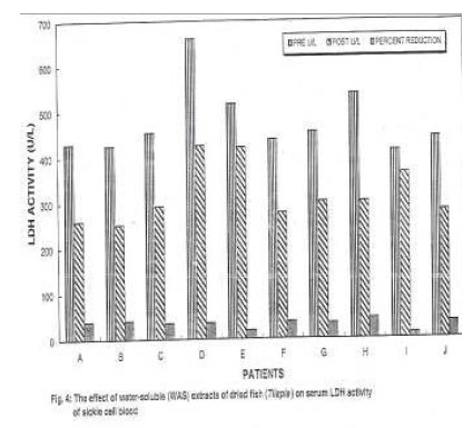
TABLE 5.a: in-vitro effect of the water-soluble (was) phase of dried fish on serum ldh activity of sickle cell blood (hbss) at a final assay concentration of 185μm l- phenylalanine equivalence
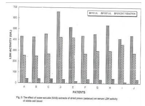
t = 5.096 tα = 1.734 TABLE 5.b: In-Vitro Effect Of The Water-Soluble (Was) Phase Of Dried Prawn On Serum Ldh Activity Of Sickle Cell Blood (Hbss) At A Final Assay Concentration Of 185μM L – Phenylalanine Equivalence
From the statistical analysis at 95% confidence level or 5% level of significance, t = 5.03; tα = 1.734; there is significant difference between the results or activities of LDH before and after the addition of antisickling agents in sickle cell blood. TABLE 6: In-vitro effect of water-soluble (was) extracts of dried tilapia and prawn on the fe2+/fe3+ ratio on sickle cell blood at a concentration of 2.07 x 10⁻⁶m phenylalanine equivalence
DISCUSSION The antisickling role of some amino acids had already been investigated and documented (Nwaoguikpe et al 1993; Ekeke and Shode; 1990). The preponderance of antisickling amino acids in the extracts of the fish and crayfishsamples must have been responsible for their profound antisickling effectiveness. The results from the hemoglobin polymerization experiments showed that the FAS, BUS and WAS fractions of the samples inhibited polymerization of HbSS very remarkably, even at very low concentrations of 500μM – Phe equivalence. Moreover, the WAS extracts were equally able to improve the Fe2+/Fe2+ ratio, hence, increasing the oxygen affinity of the sickle cell hemoglobin. One can rightly conclude that the extracts can stabilize the erythrocyte by reducing the fragility of the red cells. Consequently, these extracts (WAS) were able to reduce the activity of lactate dehydrogenase (LDH) in serum of ten sickle cell patients. This nonetheless determines the potentiality of the samples to control or reduce hemolysis of the red cells. Nutritionally, there is high quantity of free amino acids and the soluble protein concentrations of 287.3mg/100g and 1626mg/100g for Tilapia and Prawn respectively, can be of profound benefit to the sickler. We sincerely believe that dried fish (Tilapia), dried crayfish (Astacus red) and other families of fish, legumes and edible fruits can in the near future prove to be effective in the management of sickle cell disease (SCD). REFERENCES
Copyright 2005 - Journal of Applied Sciences & Environmental Management The following images related to this document are available:Photo images[ja05070f5.jpg] [ja05070f2.jpg] [ja05070f1.jpg] [ja05070f3.jpg] [ja05070f4.jpg] | |||||||||||||||||||||||||||||||||||||||||||||||||||||||||||||||||||||||||||||||||||||||||||||||||||||||||||||||||||||||||||||||||||||||||||||||||||||||||||||||||||||||||||||||||||||||||||||||||||||||||||||||||||||||||||||||||||||||||||||
| |||||||||