 |
| About Bioline | All Journals | Testimonials | Membership | News |
|
||||||
|
||||||
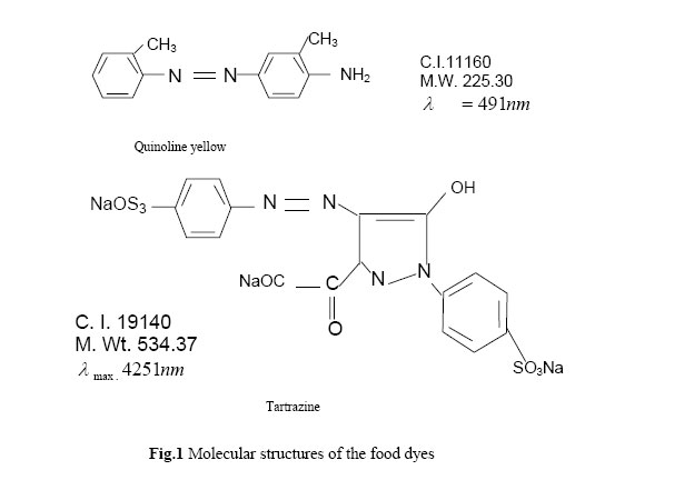
Journal of Applied Sciences & Environmental Management, Vol. 10, No. 2, 2005, pp.159-163 Biotransformation of Food Dyes by Human Intestinal Bacteria (Streptococcus faecalis, Eschericia coli) ORANUSI, N A*; NJOKU, H O Department of Microbiology, University of Port Harcourt, Port Harcourt. Nigeria. Code Number: ja06042 ABSTRACT Biotransformation of food dyes (Tartrazine and Quinoline yellow) by Streptococcus faecalis and Escherichia coli isolated from human intestinal microflora was investigated. Decolourisation of the media containing the dyes was used as an index of biotransformation. Biotransformation was higher under aerobic than under anaerobic conditions. The results obtained were attributed to the organisms cytosolic flavin-dependent reductases and redox equivalents generated by metabolism of soluble starch which transfer electrons to the chromophoric group of the dyes. The potential health risk of the resulting colourless metabolites (aromatic amines) is under investigation. @JASEM Azo dyes consist of a diazotised amine coupled to an amine or a phenol and contain one or more azo bonds (Chen et al; 1999). Azo dyes constitute about 60 – 70%of total consumption of dyes (Cartiell et al., 1995), which implies their wide occurrence in dye wastewaters. Estimates of azo dyes currently in use vary between 2,000 – 3,000 (Chen et al., 1999; Stolz, 2001) and are extensively used in the food, cosmetics, pharmaceutical industries as colourants or for dyeing in the textile and leather industries (Chen et al., 1999). The extent of their use in these industries is related to the degree of industrialisation (Chung et al., 1978). Some azo dyes and/or their intermediate metabolites used during synthesis or as by-products of microbial degradation are potentially carcinogenic, mutagenic and toxic (IARC, 1982; Houk et al., 1999). The importation of textiles, leather and other items dyed with azo dyes into Germany has recently been banned by the German Government based on carcinogenic amines (Padmavathy et al., 2003). Microbial decolourisation of azo dyes have been reported (Chen et al., 1999; Oforka and Oranusi, 2004; Cartiell et al., 1995; Oranusi and Ogugbue, 2001). This study is part of our on-going investigation on the biodegradability of food dyes (colourants) commonly used in Nigeria. This report is primarily focussed on: (i)The potential of human intestinal microflora (Escherica coli and Streptococcus facalis) to transform (decolourise) selected food dyes since it may be important in generating carcinogenic, mutagenic and toxic compounds (ii) Decolourisation of the dyes in simulated dye wastewater effluent to assess the potential of these organisms either under monoculture or consortium cultivation in the development of a bioprocess for treatment of dye wastewaters. MATERIALS AND METHODS Dyes:The azo-based food dyes were: Quinoline yellow and Tartrazine are all products of Aromco, England. They are commonly used food colourants in Nigeria. The dyes were purchased as liquid formulations. Figure 1shows the chemical structures of the dyes. Stock solutions of the dyes were sterilized by membrane filteration (pore size 0.2 μ m, Acrodisc). Media:Medium A (MA) contained in gL-1: bacteriological peptone, 10.0; meat extract, 10.0; NaCl, 5.0 and distilled water, 1,000ml Medium B (MB) is a modification of simulated dye wastewater effluent (0’Neil, 2000). The basal medium contained (gL-1): (NH4)2 SO4, 0.28; NH4Cl, 0.23; KH2PO4, 0.067; MgSO4. 7H2O, 0.04; yeast extract 0.2, NaCl, 0.15; soluble starch, 1.0, and 1ml of trace element solution containing (gL-1): FeSO4.7H2O, 5.0; ZnSO4. 7H2O, 0.01; MnCl2. 4H2O, 0.1; CuSO4.5H2O, 0.392; Co(NO3)2, 0.248; NaB4O7.10H2O,0.177, NiCl2. 6H2O, 0.025 and distilled water, 1,000 ml. MA and MB media were supplemented with appropriate sterile dye solution after sterilization and coded: MAQ or MBQ contained quinoline yellow; MAT or MBT contained tartrazine. Organism: Eschericia coli (E. coli) were obtained from the Culture Collection, Department of Microbiology, University of Port Harcourt, Nigeria. It was originally isolated from human intestinal flora and decolourised Ponceau 4R and Carmoisine (Oforka and Oranusi, 2004). Isolation and identification of Streptococcus faecalis (S. faecalis) are described below.Isolation: Ten grams of freshly voided human faeces was inoculated into duplicate 250 ml Erlenmenyer flasks each containing 100ml maltose-azide broth. Incubation was at 35+2°C with shaking at 150rpm for 3 days. After incubation, one mililitre of culture was inoculated onto series of maltose – azide agar plates and incubated as above for 48h. Colonies which were dark red or with pink centered area were picked and Gram stained. Four isolates (A, B, C and D) which were Gram positive cocci were purified by repeated plating. Stock cultures were preserved on agar slants at 40C. Standard Inoculum: Colonies were picked from the stock culture of each isolate and E. coli and inoculated into 20 ml nutrient broth contained in 250 ml Erlenmeyer flask. Incubation was at 35+2°C for 24h. Table1. Decolourisation of media which contained the dyes by batch cultures of E. coli and the isolates. Dyes
Key: +++, intense decolourisation; ++, moderate decolourisation; +, slight decolourisation; -, no decolourisation. Evaluation for Decolourisation of the dyes: One mililitre of standard inoculum of the isolates (A, B, C or D) and E. coli was each inoculated into four 25O ml Erlenmeyer flask which contained 25 ml of MA medium supplemented with 0.5ml sterile solution of each dye. Controls consisted of uninoculated flasks. Incubation was at 35+2°C with shaking at 150 rpm for aerobic cultivation. Anaerobic cultivation was in anaerobic jar (Oxoid, Hampshire, England). Cultures and controls were observed daily for growth and decolourisation (biotransformation). Based on the results obtained, E. coli and isolate B were selected Identification: Isolate B was identified using various tests: Gram stain, motility, spore stain, and various biochemical tests according to the methods of Cruickshank et al. (1980) and Holts and Bergey (1994) Isolate B was tentatively identified as Streptococcus faecalis (S. faecalis) based on the results of the various tests and with reference to Bergey’s Manual of Determinative Bacteriology. Growth and Decolourisation in Batch CultureMA Medium: Ten millilitres of standard inoculum of either E. coli or S. faecalis was inoculated into each of four 250 ml Erlenmeyer flasks, which contained 100 ml of appropriate medium (MAQ or MAT). Duplicate flasks were incubated with shaking at 150rpm at 35+2°C for aerobic cultivation while, the other two flasks were incubated under anaerobic conditions (Oxoid, Hamp shive, England). Controls for each incubation condition consisted of uninoculated flasks. Prior to and after incubation, samples (6 ml) were withdrawn for each flask for determination of percentage decolourisation. The method was as previously described (Oranusi and Ogugbue, 2001) except that the optical density was measured at λ for each dye. Table 2: Gram stain, spore test, motility and biochemical characteristics of isolate A
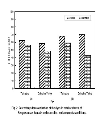
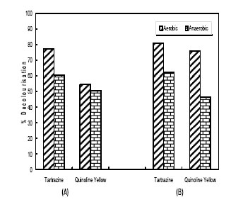
MB medium: E. coli and S. faecalisare present in aquatic environments from faecal pollution. The dyes are environmental pollutants from dye wastewaters. The potential of these organisms to transform the dyes in simulated dye wastewater effluent medium was investigated. The procedure was same as for MA medium above except that the media used were MBQ or MBT. Each medium type for anaerobic cultivation contained 100mg Na2S.10H2O per litre to remove dissolved oxygen. Incubation was at 28+2°C RESULTS AND DISCUSSIONTable 1 show the results obtained on the decolourisation potential of the isolates and E. coli after 5 days of incubation at 35+2°C. Intense decolourisation was observed in the culture of isolate B for all four dyes and in E. coli culture which contained tartrazine or quinoline yellow. Isolate A did not decolourise the dyes. Moderate and slight decolourisation was obtained in the culture of isolate C in the medium which contained quinoline yellow and tartrazine respectively. Slight decolourisation was obtained in the medium, which contained quinonine yellow and tartrazine, when inoculated with isolate D. No decolourisation was observed in the control flasks. The results of the various tests performed to identify isolate B are presented in Table 2. Isolate B was tentatively identified as Streptococcus faecalis (S. faecalis). The data obtained on decolourisation of the media which contained the dyes in batch cultures of S. faecalis and E. coli are depicited in Figs. 2 and 3. All the dye substrates were decolourised (transformed). Decolourisation efficiency was higher under aerobic conditions compared to anaerobic cultivation conditions. When S. faecalis was cultured in MAT medium percentage decolourisation was 62.40% under aerobic conditions and 56.50% under anaerobic conditions (Fig. 2A). When E. coli was cultured in MAT medium which contained tartrazine the data obtained were 77.20% under aerobic condition and 60.30% under anaerobic condition (Fig. 3A). The same trend of higher percentage decolourisation under aerobic condition than under anaerobic condition was obtained when these organisms were grown in simulated dye wastewater effluent media (MB). For example, data obtained were 68.30% and 59.30% under aerobic and anaerobic conditions respectively when S. faecalis was grown in MB medium which contained tartrazine (Fig. 2B). Corresponding data when E. coli was cultured in same medium were 80.70% and 62.40% under aerobic and anaerobic conditions respectively (Fig. 3B). There was no decolourisation in the control flasks. This showed that decolourisation was due to metabolic activities of the organisms. The sequential selective enrichment isolation procedure by culturing first on maltose – azide broth and then on medium which contained the dyes ensured the isolation of not only Streptococcus spp. but also those species, which decolourised the dyes. Reductive cleavage of the chromophoric group of dyes generates colourless intermediate metabolites (0’Neil et al., 2000). The results obtained show that the chromophoric group of the dyes was cleaved. Reduction is mediated by electron donors that transfer electrons to the chromophoric group. Electron donors may be endogenous or added into the culture medium (Russ et al., 2000; Wong and Yu, 1999). In cultures without external electron donors (Figs. 2A and 3A) the cytosolic flavin – dependent reductases acted as electron donors which decolourised the dyes. In the simulated dye wastewater effluent medium (Figs. 2B and 3B) in addition to the cytosolic flavin-dependent reductases, the metabolism of soluble starch in the medium generated redox equivalents – NADH and NADPH (electron donors). This may partially explain the higher percentage decolourisation obtained in these cultures compared to data in F Figs 2A and 3B. The enhancement of decolourisation of dyes as a result of metabolism of glucose, soluble starch and sucrose which generated external electron donors was reported by Padmavathy et al., (2003) and Oranusi and Ogugbue (2005). Bacterial azoreductases may be oxygen – sensitive or oxygen – insensitive (Shaul et al., 1991). The results obtained show that the azoreductases of S. faecalis and E. coli are oxygen – insensitive. This may partially explain the decolourisation in both aerobic and anaerobic conditions. This study has demonstrated: (i)That human intestinal bacteria (E. coli and Streptococus faecalis) decolourised (transformed) the tested food dyes under aerobic and anaerobic conditions. (ii)decolourisation in simulated dye wastewater shows the potential of these organisms to degrade these dyes in ecosystems contaminated by the dyes. We are continuing our studies on the identification of and the potential health risk of the aromatic amine generated by the microbial decolourisation of the dyes. Acknowledgement This work was funded by Senate Research Grant, University of Part Harcourt, Port Harcourt, Nigeria. We express our appreciation to C. J. Ogubue and A. Sede for technical assistance. REFERENCES
Copyright 2006 - Journal of Applied Sciences & Environmental Management The following images related to this document are available:Photo images[ja06042f2.jpg] [ja06042t1.jpg] [ja06042f1.jpg] [ja06042f3.jpg] [ja06042t2.jpg] |
| |||||||||