 |
| About Bioline | All Journals | Testimonials | Membership | News |
|
||||||
|
||||||
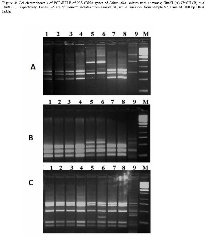
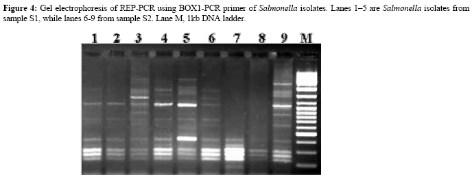
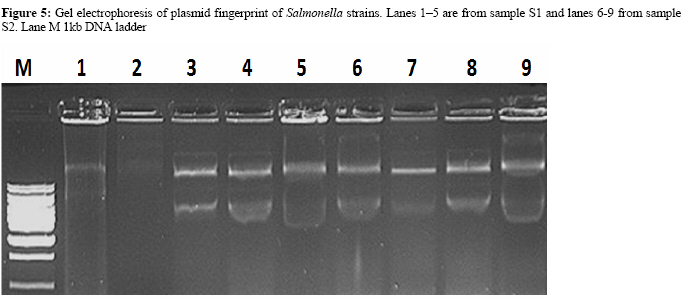
Molecular and biochemical diagnosis of Salmonella in wastewater 1Sahar, Zaki; 1*Desouky, Abd-El-Haleem; 2Ehab, El-Helow; 1Marwa, Mustafa 1Environmental Biotechnology Department, Genetic engineering and Biotechnology Research Institute, Mubarak City for Scientific
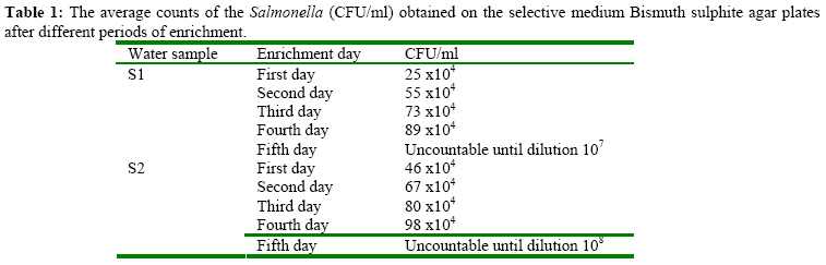
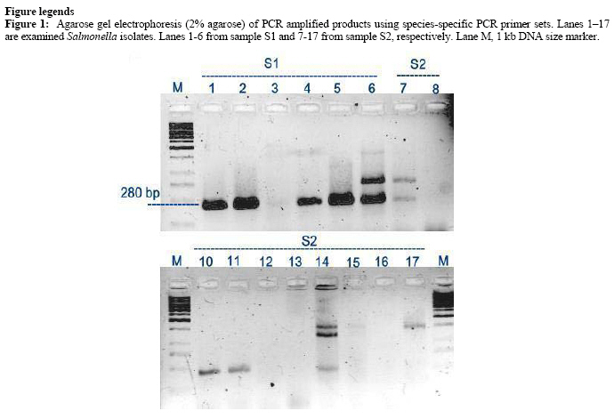
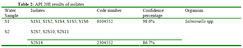
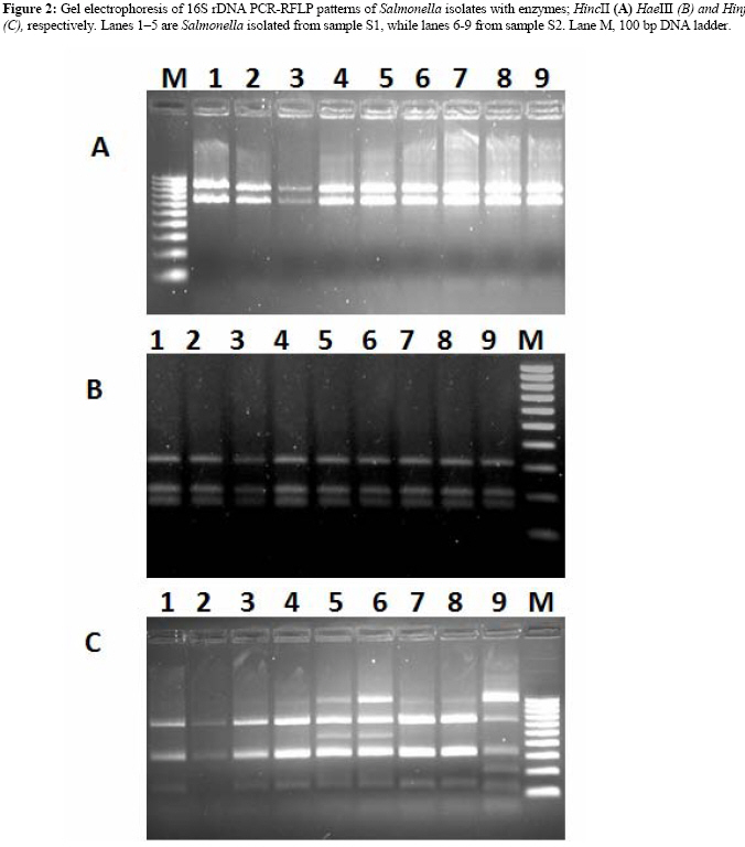
research and Technology Applications, Burgelarab City, Alexandria, Egypt Code Number: ja09029 ABSTRACT This study aimed to employ biochemical and molecular assays to detect and diagnose Salmonella in wastewater. For this reason, two water samples were collected from Alexandria wastewater treatment plant (S1) and septic tank of a hospital at Alexandria governorate (S2). Selective culture media specific for Salmonella were used to grow and purify a number of isolates from water samples. Direct plate count revealed a high frequency of Salmonella cells in sample S2 than S1. As a confirmatory, species-specific PCR assay was performed for 17 randomly selected bacterial isolates from both water samples. Positive-PCR Salmonella isolates (5 from S1 and 4 from S2) were subjected for identification using API 20E biochemical identification kit. Among various examined antibiotics, rifamycine was the most effective antimicrobial agent on Salmonella isolates. Subsequently, PCR-RFLP of 16S and 23S rDNA genes, Rep-PCR fingerprinting and plasmid profile were employed to recognize among isolates. Out of nine examined restriction endonucleases, HinfI, HincII and HaeIII were the most discriminative enzymes which allowed clear differentiation among isolates, while PCR-RFLP of 23S rDNA was most discriminative than 16S rDNA. Higher levels of polymorphism and specificity were achieved by reproducible genomic fingerprints of Rep-PCR. However, poor discrimination results were achieved by employing plasmid profile assay. @ JASEM Salmonella is one of the most important pathogens involved in human food-borne illness (Oliveira et al., 2003). The majority of human salmonellosis cases are caused by the consumption of contaminated egg, poultry, pork, beef and milk products (Geimba et al., 2004). Salmonella causes a serious health problem in developing countries through a wide range of human diseases such as enteric fever, gastroenteritis and bacteremia (Banavandi et al., 2005). The infective dose of Salmonella can be as low as 15 to 20 cells, depending upon age and health of the host (FDA, 2003). The increase of pollution in natural water has intensified the detection frequency and persistence of pathogenic microorganisms mainly Salmonella in areas affected by sewage discharge. Salmonella infection of humans is a worldwide health problem through water consumption (Arvanitidou et al., 1995). However, contaminated water can also be a major factor in the spread of epidemics (Clark et al., 1996). It is established that, the microbiological quality of water is based on the detection of total coliform and faecal streptococci as indicator organisms. In 1976, European Economic Community (EEC) reported that Salmonella must be checked in addition to the indicators. Authors have claimed that intermittent presence of Salmonella has been seen as a result of concrete case contamination (Moriñigo et al., 1993). Traditional detection methods for Salmonella are based on cultures using selective media and followed by a series of biochemical and/or serological tests. Generally, 5-7 days are required to confirm the presence of Salmonella species (Kumar et al., 2003). Detection of Salmonella enterica in faecal or water samples is limited due to low numbers of the bacterium, thus necessitating the use of an enrichment step. Enrichment and pre-enrichment broths can dilute inhibitory compounds produced by competing bacteria in the sample, as well as aid in recovery of stressed cells. Generally, isolation of Salmonella from samples, such as faeces, that contain >107 aerobic bacterial cells/g requires a selective enrichment medium that permits the growth of Salmonella, while inhibiting the growth of other aerobic bacteria (Madigan et al., 1997). However, some selective enrichment media also inhibit the growth of Salmonella cells that are stressed or damaged (Chen et al. 1993). In order to overcome the above drawbacks, PCR has been tested for the rapid detection of bacterial pathogens in a number of recent studies. It has been applied in two distinct ways; i) the rapid identification of pathogenic bacteria species which had been isolated from environmental sources; examples include species of Bifidobacterium (Kaufmann et al., 1997), Staphylococcus and Salmonella (Abd-El-Haleem et al., 2003), Listeria and E. coli (Osek, 2002), and P. aeruginosa (De-Vos et al., 1997) and ii) the direct detection of bacterial pathogens in environmental samples without the requirement for isolation on artificial media (Abd-El-Haleem et al., 2003). Like bacteriological assays, PCR often requires enrichment of fecal samples (non-selective and selective enrichment) to increase Salmonella numbers, improve PCR sensitivity and to aid in the dilution of compounds that may interfere with the PCR (Oliveira et al., 2003). Several sets of primers designed to detect genes specific for Salmonella. These included hilA (hyperinvasive locus A, a gene that encodes one of the proteins required for the regulation of virulence genes) in Salmonella in different samples (Lesnick et al., 2001); ompC (encodes a major structural outer membrane protein) been proposed as another target for detection of Salmonella in clinical samples (Amavisit et al., 2001); and invA, (essential for full virulence in Salmonella and thought to trigger the internalization required for invasion of deeper tissues) and thought to be specific for Salmonella (Castagna et al., 2005) The purpose of the present study was to evaluate the utility of biochemical assays, species-specific PCR, PCR-RFLP fingerprinting of ribosomal DNA (16S and 23S rDNA genes), Rep-PCR and plasmid profile for identification and typing of Salmonella isolates from two wastewater sources. MATERIALS AND METHODSWater samplesSewage water samples were collected from two different sources in the governorate of Alexandria. These included the West-Alexandria wastewater treatment plant (before treatment) (designated S1) and the septic tank of El-Omal hospital (designated S2). Water samples were collected in triplicates according to the Standard Methods for the Examination of Water and Wastewater (APHA 1995). Two liters sterile plastic bottles were used. Subsequently, water samples were transferred in an ice box to the laboratory within 6 h of collection for bacteriological analyses. Isolation and enumeration of salmonella In all cases, water samples were shaken vigorously, and dilutions were prepared in sterile saline (0.9 g NaCl/100ml of sterile distilled water). Triplicate plates were used for each sample dilution. The original numbers of organisms (CFU/ml) in water samples were calculated considering the dilution factor and the final estimate was taken as the average of figures obtained from the countable plates (APHA 1995). Subsequently, isolation of Salmonella was performed by adding an aliquot of 0.1 ml from the water sample to 4.9 ml of Tetrathionate broth medium for enrichment and incubated at 35˚C for 5 days. During the 5 days, repeated spreading was made from the same broth medium after each 24 h period onto bismuth sulfite agar medium. Plates were incubated at 35˚C for 24 h at inverted position. Typical colonies were developed with a black color (APHA, 1995). For maintaining, isolates were preserved by freezing at -80˚C in nutrient broth medium containing 10% glycerol (Berardesco et al., 1998). In addition, bacterial isolates were maintained, for short periods of time by continuous sub-culturing on nutrient agar and keeping in a refrigerator. Biochemical diagnosis of Salmonella The Analytical Profile Index (API20E strips) was used as a biochemical system for identification of Salmonella. The API20E strip consists of 20 microtubes containing dehydrated substrates. These strips were incubated in bacterial suspensions for 18 to 24 h at 37˚C. During the incubation period, metabolism produces changes that are either spontaneous or revealed by the addition of reagents. The standard was scored according to a reading table and the identification was obtained by referring to the API catalogue (Clayton, 1999). Furthermore, the sensitivity of the isolates to antibiotics was examined by streaking on LB-agar plates, each containing one of the following antibiotics: ampicillin (50 mg/l), kanamycine (50 mg/l), tetracycline (10 mg/l), erythromycin (50 mg/l), penicillin 50mg/l, chloramphenicol (50 mg/l) Neomycin (50 mg/l), streptomycin (50 mg/l), and rifamycine (50 mg/l). Molecular diagnosis of Salmonella Species-specific PCR In all cases, DNA extraction and purification from both water samples and bacterial isolates were performed according to that previously described by Abd-El-Haleem et. al. (2003). As described by Carli et. al. (2001), primer pair, highly specific for particular target sequences invA was used to test for Salmonella. The sensitivity and specificity of the PCR reactions were also optimized as described elsewhere (Abd-El-Haleem et al., 2003). PCR-RFLP fingerprint of 16S and 23S rDNA genes PCR-RFLP of amplified 16S and 23S rDNA genes of reference stains and isolates was performed. Amplification of 16S and 23S rDNA fragments of ~1.3 and 3 Kb in size with universal eubacterial PCR primers was carried out as described previously by Abd-El-Haleem et. al. (2002). Subsequently, the PCR amplified products were digested with 2 U of restriction enzymes SacI, NotI, BamHI, XbaI, SpeI, SauIII, EcoRI, HindIII, HaeIII, HincII and HinfI (GIBCO B.R.L) according to the recommendations of the manufacturers and electrophoresed in 2% agarose gel in the presence of ethidium bromide. Gels were run at 100 V for 3 h in 1XTBE buffer and then visualized and photographed in the MultiImage light cabinet (Alpha Innotech Corporation, USA). 100 bp and 1 kb ladder mix (Sigma, St. Louis, MO) were used as molecular weight markers. Rep-PCR Amplification of repetitive sequences that are scattered along DNA molecules was carried out using the PCR primer BOX1 (Louws et al., 1994). The PCR mixture consisted of 5 µl of 10×PCR buffer, 3 µl MgCl2 (50 mM),4 µl of dNTPs (2.5 mM each), 1 µl of primer (10 pmol/µl), 0.5 µl of 5 U/µl Taq DNA polymerase, 1.5 µl of the isolates DNA (50 to 100 ng/µl), and 34 µl of distilled water. PCR amplification was stared with 94°C for 5 min, followed by 30 cycles of 92°C for 45 sec, 1 min at 50°C, and 6 min at 65°C. After amplification, there was an additional extension step at 65°C for 7 min, and then the samples were cooled to 4°C. Each PCR mixture was electrophoresed in a 2% agarose gel containing ethidium bromide at 0.1 µg/ml. Gels were run at 6 V/cm for 120 min in 1X TBE and PCR products were visualized and photographed in MultiImage light cabinet (Alpha Innotech Corporation, USA). Gene ruler ladder mixes (Fermentas) were used as molecular weight markers. Plasmid profile Plasmid DNA was isolated by the procedures of Connors and Barnsley (Connors and Barnsley, 1982). Plasmid profiles were visualized by ethidium bromide staining of 0.8% (w/v) agarose gels. RESULTS Isolation, identification and molecular confirmation of Salmonella Because of the high solid contents of sewage and the anticipated low densities of Salmonella in both water samples, an enrichment procedure was carried out to increase bacterial growth. Plate counting results after 1 to 5 days of enrichment are listed in Table 1. After their growth on specific media, randomly selected 6 isolates from sample S1 and 11 isolates from sample S2 were subjected for molecular conformation using species-specific PCR primers (Figure 1). Positive PCR isolates were maintained for subsequent phenotypic and fingerprinting diagnostic examinations. The specificity and sensitivity of the primers were examined against the same reference strains described previously by Abd-El-Haleem et. al. (2003). Subsequently, the nine PCR-positive isolates (5 from sample S1 and 4 from sample S2) were subjected for biochemical identification using the API 20E strips. The results demonstrated a correct genus identification of examined Salmonella isolates. Nevertheless, variations in the identification confidence (expressed as a percentage) are listed in Table 2. For further recognition among Salmonella, isolates were subjected to a number of antibiotic sensitivity assays. Nine different antimicrobial agents, representing six different groups of antibiotics, were used. The results demonstrated that all isolates are resistant to penicillin and erythromycine while rifamycine was the most effective antimicrobial agent used. Out of these data, it could emphasize that ampicillin, neomycine, kanamycine, tetracycline, chloramphenicol, and rifamycine can serve as tools of discrimination among members of Salmonella. However, the pattern of sensitivity of Salmonella isolates to the nine examined antimicrobial agents was as follow; rifamycine > chloramphenicol > neomycin, kanamycine > tetracycline, streptomycin > penicillin and erythromycin, respectively. PCR-RFLP of 16S and 23S rDNA genes All API20E identified isolates were subjected for analysis by restriction fragment length polymorphisms (PCR-RFLP) of the 16S and 23S rDNA genes, while several restriction endonucleases were separately used to digest the PCR products. PCR amplification of ribosomal DNA genes was carried out using universal primers described in the materials and methods. Negative controls (PCR reaction without DNA templates) revealed no amplification products (data not shown). PCR-RFLP analysis of 16S RDNA genes was performed using each of the following restriction endonucleases: SacI, NotI, BamHI , XbaI, SpeI, SauIII, EcoRI, HindIII, HaeIII, HincII and HinfI. In cases of NotI, BamHI, XbaI, SpeI and SauIII, the results showed poor digestion patterns with all isolates. However, despite a good digestion occurred with the endonucleases HeaIII, HincII, SacI, EcoRI, and HindIII, the resulted banding patterns didn't reveal satisfactory discriminations between isolates. Figure 2 A and B show the fingerprint patterns of the enzymes HeaII and HincII, respectively. On the other hand, reasonable digestion results and reproducible fingerprint patterns that can be interpreted easily by the naked eyes were observed with enzyme HinfI (Figure 2C). The genotyping pattern of Salmonella strains with enzyme HinfI demonstrated that they can be classified into three groups. The first includes isolates S1S1, S1S2, S1S3, S1S4, S2S10 and S2S11. However, the second includes isolates S1S5 and S2S7. While the third contains only isolate S2S14. In contrast to PCR-RFLP of 16S rDNA amplified genes, the best discriminatory patterns of 23S rDNA genes were obtained with three enzymes HincII, HeaIII and HinfI. As shown in Figure 3A-C, HincII was able to discriminate salmonella isolates into five groups with a common band at 780 bp. These include S1S1 and S1S2, (group1), S1S3 and S1S5 (group 2), S1S4, S2S10 and S2S11 (group 3), S2S7 (group 4) and S2S14 (group5). However, resulted HaeIII banding patterns were able to differentiate isolates into three groups; the first contains S1S1, S1S2, S1S3, S1S4, S2S10 and S2S11, the second include strains S1S5 and S2S7, while the third include only strain S2S14. HinfI discriminate Salmonella strains into three recognized groups including; S1S1, S1S2, S1S3, S1S4, S1S5 S2S10 and S2S11 (group 1), S2S7 (Group 2) and S2S14 (group 3). Rep-PCR As shown in Figure 4, Rep-PCR profiles were able to discriminate clearly among isolates. The number of bands per each profile varied within the range 4-14 with variable intensities and apparent molecular weights ranging from 200 bp to 3500 bp. Four common bands at 220, 250, 300 and 400 bp were detected in all Salmonella strains. Plasmid profiling Examined Salmonella isolates were compared with respect to their plasmid profiling. As shown in Figure 5, all Salmonella isolates are containing plasmids. DISCUSSION In contrast to traditional methods, PCR-based techniques can be applied to detect culturable and non-culturable Salmonella in water within hours instead of days required in conventional biochemical assays (Abd-El-Haleem et al., 2003). In the present study, water samples had been collected from Alexandria wastewater treatment plant (before treatment), designated S1 and septic tank of El-Omal hospital, designated S2. However, quantification of Salmonella was depending on the growth of bacteria represented by a single cell to a visible colony on a solid medium. The results demonstrated that the count of Salmonella were higher in hospital wastewater sample S2 than S1. Subsequently, 17 single colonies of Salmonella grown on their specific selective media were randomly selected and subjected to molecular conformation using species-specific PCR primers. Positive-PCR Salmonella isolates (5 from S1 and 4 from S2) were then subjected for identification using API20E biochemical identification kit which exhibits a high degree of reproducibility. Identified Salmonella isolates were examined for their antibiotics sensitivity. All of them showed 100% resistance to penicillin and erythromycin, which belong to the β-lactams and macrolides groups, respectively. It has been previously reported that Salmonella are greatly resistant to ampicillin, penicillin and erythromycin (Ehinmidu, 2003). However, rifamycine is appeared to be the most effective antibiotic. Rifamycine causes inhibition of transcription; while the strains that resist rifamycine altered DNA dependent RNA polymerases preventing binding of the antibiotic (Yee et al., 1996). However, multiple resistances to antibiotics were commonly observed in all strains which could be, at least partially, a result of the presence of plasmids or due to the combination of several mechanisms (Seepersadsingh et al., 2005). Then, the nine salmonella isolates were subjected to PCR-RFLP analysis of 16S rDNA genes. The results allowed obvious discrimination between the studied isolates at the genus level. In agreement with Abd-El- Haleem et. al. (2003), HinfI was the most suitable enzyme for RFLP analysis of the 16S rDNA amplicons. It allowed obvious discrimination by producing interpretable PCR-RFLP patterns. The exceptional HinfI banding pattern of Salmonella S2S14 indicated that it is a unique strain. Historically, the 16S rDNA gene has been chosen by many researchers for phylogenetic studies due to its relatively small size which facilitates sequence analysis. However, for its sequence similarity among and within some microbial species, 16S rDNA sequences may be insufficient (Abd-El-Haleem et al., 2002). Under such circumstances, 23S rDNA sequences become more attractive. Being twice (about 3 kb) as long as the 16S rRNA, it contains more genetic information and more variable regions (Ludwig and Schleifer, 1999). For instance, the 23S rDNA gene has been used for identification of different microbial species (Redwan and Abd-El-Haleem, 2005). In contrast to the 16S rDNA PCR-RFLP in our study, three enzymes HincII, HeaIII and HinfI were the most discriminative restriction endonucleases and produced different 23S rDNA polymorphic patterns among Salmonella isolates. Our results demonstrated that 23S rDNA gene contains more restriction sites than 16S rDNA which allow more discriminatory power. Previously, RFLP of the 23S rDNA genes has been used for determination of the genetic heterogeneity among the isolates from contaminated buffers during diagnosis of HCV infections (Redwan and Abd-El-Haleem, 2005), identification of Campylobacter and Arcobacter species (Hurtado and Owen, 1997) and phylogeny of Rhizobium galegae (Terefework et al., 1998). The results presented in this study, suggested that amplification of 23S rDNA genes followed by RFLP analysis with one of restriction endonucleases HaeIII, HincII and HinfI can be applied for typing Salmonella after isolation by classical microbiological methods. This would allow confirmation and strain differentiation as well, avoiding the time consuming biochemical and serological confirmatory assays. Rep-PCR methods have now been widely applied in the research of bacterial diversity and fingerprinting of clinical isolates as well as strains of industrial, agricultural and environmental organisms (Woo and Lee, 2006). In the present work, reproducible genomic fingerprints with high levels of polymorphism and specificity were produced when Rep-PCR was performed using the BOX1 primer. Interestingly, the Rep-PCR profiles were able to clearly discriminate all of examined Salmonella isolates confirming that each strain has a unique distinguishable genotype. Such a high discriminatory power of Rep-PCR has been previously reported elsewhere (Woo and Lee, 2006). Plasmid profiling is a relatively simple and rapid technique and can be useful for laboratories that are unable to perform more complex methods (Liebana, 2002). Some studies have been shown that certain plasmid profiles are stable among Salmonella isolates even when isolated from cases of infection occurring in widely separated geographic areas (Chadfield et al., 2001). This method was also sensitive when applied for typing of S. typhimurium, S. virchow and S. gallinarum strains (Rasschaert et al., 2005). However, plasmid analysis was not found to be suitable for typing of strains of S. enteritidis (Domig et al., 2003). In the present study, plasmid profile genotyping failed to discriminate between salmonella strains as eight out of nine have almost identical sizes and number. In fact, plasmid profile should be used in combination with, but not in isolation from, other epidemiological methods (Açik et al., 2005). Finally, this study recommends use of PCR-based methods followed by RFLP analysis of ribosomal RNA genes as rapid, sensitive, specific and accurate technique for detection and typing of Salmonella isolates. In addition these techniques can easily and efficiently discriminate between different pathogens when compared to biochemical and serological methods. ACKNOWLEDGEMENTS Genetic Engineering and Biotechnology Research Institute at Mubarak city for Scientific Research and Technology Applications supported this work. REFERENCES
Copyright 2009 - Journal of Applied Sciences & Environmental Management The following images related to this document are available:Photo images[ja09029t2.jpg] [ja09029f3.jpg] [ja09029f5.jpg] [ja09029f4.jpg] [ja09029f2.jpg] [ja09029f1.jpg] [ja09029t1.jpg] |
| |||||||||
