 |
| About Bioline | All Journals | Testimonials | Membership | News |
|
||||||
|
||||||
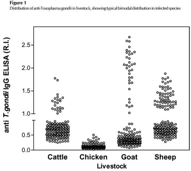
African Journal of Health Sciences, Vol. 13, No. 1-2, Jan-June, 2006, pp. 131-134 Serological survey of antibodies to Toxoplasma gondii* Ciamak Ghazaei Mohaghegh Ardabili University ; Iran-Ardabil-Phase 1 Sabalan-Plake 722 Tel: +98- 0452- 7463418; Fax: +98- 0452- 7463417 Code Number: jh06018 SUMMARY Toxoplasmosis is one of the most prevalent parasitic infections of man and livestock, and its transmission has usually been attributed to ingestion of undercooked or raw meat from infected livestock, with the infection rate in those animals being an important risk predictor of human disease, high in Iran and Ardabil State. During a study on this public health problem, we tested serum samples from cattle, goats, sheep and chicken from the State of Ardabil, Iran, for IgG antibodies to Toxoplasma gondii by enzyme-linked immunosorbent assay (ELISA). Antibodies to Toxoplasma gondii were found in 30% (60/200) of sheep, 15% (30/200) of goats and 9% (18/200) of cattle, and none were found in chicken sera. Despite the differences in feeding habits of each species, the rate of infection of the animals tested could be attributed to livestock management methods, whose improvement could reduce infection. [Afr J Health Sci. 2006; 13:131-134] *Published online before print Introduction Infection by the protozoan parasite, Toxoplasma gondii, is widespread in humans and many other species of warm-blooded animals [1,2,3,4]. Although the course of disease is generally benign, it can cause significant morbidity and mortality in the developing fetus and in immunocompromised individuals, including humans with Acquired Immunodeficiency Syndrome (AIDS) or those submitting to cancer chemotherapy. Among livestock, sheep and goats are more widely infected with Toxoplasma gondii than cattle and chicken [2,3,4,5]. This parasite is a major cause of abortion, with significant economic losses to sheep and goat breeders. The infection does not usually cause clinical symptoms in cattle and in poultry [1, 6, 7 ]. Recent studies showed [8, 9] that a small percentage of affected individuals acquire infection in the uterus, but the majority become exposed to Toxoplasma gondii by ingestion of undercooked or raw meat containing tissue cysts, ingestion of oocysts shed by infected cats or consumption of contaminated drinking water or fresh vegetables. In Iran, Toxoplasma gondii have been found in mutton, goat meat, beef and chicken [10]. Although Toxoplasma gondii is found in most parts of the world, there have been relatively few recent reports on small ruminants in cattle and chicken in Iran [11,10]. Epidemiological surveys are the most useful way of assessing the relative importance of different sources of Toxoplasma gondii infection in humans. Since contaminated meat is a significant infection source to man, it is particularly beneficial to ensure continuous surveillance of T. gondii prevalence in animal species destined for human consumption. Materials and Methods Sera were collected from a total of 750 food animals from Ardabil State, Iran. 200 being from extensive breed cattle slaughtered at abattoirs in the city of Ardabil, 200 from semiintensive breed goats from farms in the Ardabil region, 200 from extensive breed sheep slaughtered at abattoirs in the city of Ardabil and 150 from intensive breed chicken slaughtered at abattoirs in the city of Ardabil. Cattle, sheep and chicken blood were collected during slaughter, immediately after killing, and goat blood was collected by venipuncture. Sera were separated from clot by centrifugation at 1000g for 10 min, mixed (1:1, v/v) with phosphate buffered glycerol, pH 7.2, and stored at -200C until use. RH strain Toxoplasma gondii tachyzoites salt soluble antigen was prepared from infected mouse peritoneal fluid as described elsewhere, except for one step of mammalian cell exclusion by adhesion to sterile pre-packed Sephadex G50 columns. The antigen was adjusted to 1mg protein/ml and stored at -700C until use. ELISA was performed using high protein binding-certified microplates (Sigma�) coated with 100 ml/well of Toxoplasma gondii antigen at a concentration of 10 mg/ml [12,10,11]. Each serum sample, diluted at 1:100 in PBS-T, was added to each well and bound IgG detected with speciesspecific anti IgG peroxidase conjugate, with optical density (OD) measured in a microplate reader. In each plate was included a positive, usually obtained from an experimentally infected animal from the same species, a negative and threshold controls, all previously determined by Indirect Immunofluorescence Assay (IFA). The threshold control, obtained from the dilution of the positive serum of known IFA titer in standard negative serum, was used to distinguish reactive from nonreactive serum samples in multiple plate assays; the absorbance of the threshold serum was taken as the lowest level of identifiable positive reaction. The reactivity index (RI) of the samples was defined as the ratio of the average absorbance of the samples by the average absorbance of the threshold serum, being positive when RI³ 1.0. All serum were tested in duplicate, with reproducibility inter and intra tests higher than 99.00%. Results The ELISA results were shown both as frequency of infection and also as their reactivity distribution as in Figure 1. Results showed 31.00% of seropositivity in sheep, 17.00% in goat, 11.00% in cattle, but no positive sample in poultry with a 95.00% confidence interval for each measure, which was significantly higher in sheep than in goats and cattle, and absent in the chicken. Their reactivity distribution clearly showed the expected bimodal distribution of infected and non-infected animals. Comparison between frequencies by c2 test showed that the highest frequency was found in sheep, with cattle and goats with similar intermediate. Table 1: Frequency of seropositivity for T. gondii among livestock from Ardabil state, Iran.
Discussion Serologic prevalence data indicate that Toxoplasmosis is one of the most common of human infections throughout the world. Infection is more common in warm climates and at lower altitudes than in cold climates and mountainous regions. High prevalence of infection in France has been related to a preference for eating raw or undercooked meat, while high prevalence in Central America has been related to the frequency of stray cats in a climate favoring survival of oocysts. There are two basic forms of Toxoplasma organism: the "oocyst," which is shed in the cat feces, and the Toxoplasma tissue stages, which live in the flesh of such food animals as hogs and lambs. A person who inadvertently eats either of these forms of Toxoplasma is liable to become infected. [1,2,9]. When the infected person is a pregnant woman, the Toxoplasma organism may cross the placenta. The amount of damage done to the mother and the fetus/baby depends on the stage of pregnancy at the time of infection. Infection in early pregnancy may result in miscarriage or stillbirth or in a child with varying degrees of blindness (due to inflamed retina) and/or various severe neurological conditions including hydrocephalus, microcephaly, and retardation. Sometimes problems are not evident at birth and show up later in life. There is, however, a very strong association between Toxoplasma infection and working with raw meat such as in slaughterhouses or as a butcher. The results of this survey would suggest that small ruminants play a more important role as a source of Toxoplasmosis than cattle, as discussed elsewhere. Nonetheless, despite the lower seroprevalence detected in cattle, consumption of mutton is much greater than that of beef or goat meat and thus increases the importance of sheep as a source of local infection. The current understanding of the epidemiology of Toxoplasmosis leads us to think that herbivores acquire infection by ingestion of pasture and water contaminated with Toxoplasma gondii oocysts shed by cats. The differences in rates of infection could be attributed both to differences in susceptibility to T. gondii or to differences in management methods. Since sheep are bred under extensive-management, they are more likely to be exposed to Toxoplasma gondii oocysts in pasture and water than goats, which are supplied with quality water and food under semi-intensive management. Lower seropositivity in cattle samples compared to those in sheep may be attributed to differences in susceptibility, since both species are bred under extensive management. On the other hand, the absence of infection in the chicken can also be attributed to the management method used, as these animals are bred under highly intensified management systems. The data showed that, out of the three infected species, the lowest seroprevalence occurred in cattle, but in view of the typical preference for beef, bovine protein cannot be ruled out as a significant source of human infection, including processed products. The high infection rate in sheep might have local implications because, in the State of Ardabil and Iran, mutton is more popular as a source of animal protein. This makes it a potential source of human Toxoplasmosis of increasing importance. Conclusion The data suggest that it is possible to significantly reduce the risk of Toxoplasma gondii infection in livestock using intensive farm management with adequate measures of hygiene, confinement, and prevention. Suggested precautions include:
References
Copyright 2006 - African Forum for Health Sciences The following images related to this document are available:Photo images[jh06018t1.jpg] [jh06018f1.jpg] |
| |||||||||
