 |
| About Bioline | All Journals | Testimonials | Membership | News |
|
||||||
|
||||||
Journal of Medicine and Biomedical Research, Vol. 3, No. 1, June 2004, pp.37-41 Correlation between cyanide-induced decreases in ocular Ca2+-ATPase and lenticular opacification NP Okoliea and K Audua aDepartment of Biochemistry, Faculty of Science,
University of Benin, PMB 1154, Benin City, Nigeria.
Code Number: jm04005 ABSTRACT The effect of cyanide toxicity on Ca2+-ATPase activities in lens, cornea and vitreous humour was investigated in New Zealand White rabbits fed for seven weeks on either pure growers mash or mash containing 400ppm inorganic cyanide. Results indicate that Ca2+-ATPase activities were significantly decreased in the lens and vitreous humour of the cyanide-fed rabbits (p < 0.05), while the corneal enzyme was unaffected. Ophthalmoscopic examination of the cyanide-exposed rabbits revealed adverse morphological changes, including pale fundus, low retinal reflex and lenticular opacification. The results suggest that cyanide-induced cataractogenesis may be a consequence of disruption of vitreous humour and lenticular calcium homeostasis. Key Words: Cyanide, Ca2+-ATPase, cataracts INTRODUCTION Cyanide is a potent respiratory poison in all aerobic organisms. It combines irreversibly with ferrocytochrome a/a3 thereby inhibiting electron transport, mitochondrial oxygen uptake and cellular respiration.1,2 Humans are exposed to cyanide through dietary and environmental sources.3 The dietary routes of cyanide exposure include cassava4, legumes 5 and cereal grains6; while one major environmental source of cyanide is tobacco smoke.7 Indeed it has been suggested that cyanide in tobacco smoke might be the principal culprit in the pathogenesis of tobacco amblyopia, characterised by sudden dimness of vision amongst smokers.8 However, the precise mechanism by which cyanide interferes with visual acuity remains to a large extent unclear. Recently it has been reported that cyanide imposes oxidative stress on the lens, compromises ocular antioxidant status, and triggers off lenticular opacification in rabbits.9 Studies have shown that calcium ions play a vital role in the development of cataracts.10,11 Indeed the maintenance of Ca2+ homeostasis by lenticular Ca2+ ATPase is an imperative for lens clarity.12 Inhibition of Ca2+-ATPase has been shown to cause Ca2+ accumulation and lenticular opacification.13 The present study was carried out to investigate the influence of cyanide exposure on some ocular tissue Ca2+-ATPases so as to see if a link exists between cyanide-induced visual impairment and alterations in calcium homeostasis. MATERIALS AND METHODS Animals and feeding Two-month-old New Zealand White rabbits (initial mean weight 1.072kg) were purchased from a breeder in Benin City. The animals were housed in clean rabbit hutches and acclimatised on growers mash (Bendel Feed and Flour Mills BFFM Ltd, Ewu, Nigeria) for three weeks prior to the study. They were subsequently assigned randomly to two groups (seven rabbits each). One group (test) was fed growers mash containing 400ppm cyanide. To achieve this, 1g of potassium cyanide was thoroughly mixed with 1000g of growers mash in a plastic bowl prior to presentation. Members of the second group were fed pure mash and served as controls. Before feeding, each feed type was mixed with distilled water in the ratio of 10:1 (w/v) to minimise feed dust and enhance accept-ability. Fresh feed was provided on daily basis, while stale remnants were discarded after weighing. On the average, each rabbit received 130g feed/day; and clean drinking water was provided ad lib. At the end of seven weeks, the animals were weighed and their eyes were carefully examined using an ophthalmoscope. They were subsequently sacrificed by cervical dislocation, and their eyeballs were carefully dissected out into a beaker containing physiological saline (0.9% NaCl w/v).
Preparation of tissue homogenates The lens, vitreous humour and cornea of each eyeball were dissected out. Each tissue was finely ground in a hand mortar with 1.0ml of ice-cold physiological saline (0.9% NaCl w/v) for two minutes. The homogenate was then transferred into a vial and preserved in a refrigerator at 4OC. All tissue extracts were analysed within 48 hours.
Assay of Ca2+-ATPase Ca2+-ATPase was assayed by estimating the amount of inorganic phosphate liberated following incubation of aliquots of tissue homogenate with disoduim ATP.15 The assay mixture contained 0.1ml of 1mm EDTA; 0.4ml of 30mM MgCl2; 0.4ml of 5mm CaCl2; 0.3ml of 160mM tris buffer, pH 7.4 and 0.4ml of 0.3mM disodium ATP. Reaction was started by the addition of 0.4ml tissue homogenate. The mixture was vortexed and incubated at room temperature (≈30°C) for 30 minutes, after which 4.0ml of 5% (v/v) trichloroacetic acid was added to stop the reaction. The mixture was subsequently clarified by centrifugation, and inorganic phosphate estimated in the supernatant using ammonium molybdate.16 Corrections were made for endogenous inorganic phosphate arising from inherent ATP hydrolysis by incorporating a homogenate ATP blank in the assay. CA2+-ATPase was expressed in terms of μg inorganic phosphate liberated/ml of tissue extract. Statistics Means + SEM values for parameters were compared between tests and controls using Student's t-test, and p values < 0.05 were taken as significant.
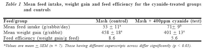
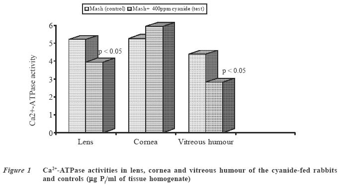
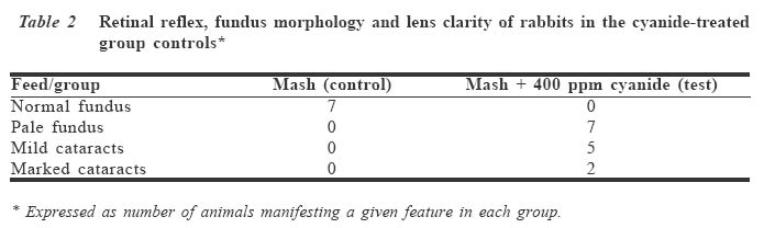
RESULTS Table 1 shows the mean feed intake, weight gain and feed efficiency values for rabbits in both groups. The cyanide-treated rabbits consumed significantly higher feed (p < 0.05) than those in controls, although this did not translate into superior weight gains for this group. Figure 1 shows the pattern of changes in Ca2+-ATPase activities in some ocular tissues as a result of cyanide exposure. The enzyme activity was significantly decreased in the lenses and vitreous humour of the cyanide-toxified rabbits (p < 0.05). However, there were no significant changes in corneal Ca2+-ATPase. Table 2 shows results obtained in ophthal-moscopic examination of rabbits in both groups before sacrifice. While the rabbits in the control group showed normal features, those in the cyanide-fed group had weak retinal reflex, pale fundus and lenticular opacification (cataracts). DISCUSSION Lenticular Ca2+-ATPase functions to maintain low internal calcium within the lens through the removal of cytosolic calcium across the plasma membrane.12 Disturbances in lenticular calcium homeostasis have been associated with opacification of the lens and cataracts.17-19 This is in agreement with results obtained in the present study, in which the cyanide-induced significant decreases in Ca2+-ATPase activities in lens and vitreous humour were accompanied by opacification of the lens. It is known that Ca2+ accumu-lation within the lens stimulates proteolysis of lens proteins, leading to deleterious changes in lens permeability, thereby making it opaque.8,20 Indeed it has been demonstrated that the activity of Ca2+-ATPase is 50% less in human cataractous lenses than in clear ones.19 Thus, the significant reductions in Ca2+-ATPase activities in the lens and vitreous humour of the cyanide-toxified rabbits are consistent with the observed functional and morphological changes in the lens, fundus and retina. The vitreous humour is a gelatinous matrix that helps to maintain the shape and pliability of the eye.12 Clarity of vitreous humour ensures minimal light scattering, and enhances vision. As a result, the cyanide-induced reduction in vitreous humour Ca2+-ATPase activity will have adverse consequences for vision, arising from alterations in its clarity and permeability due to disruption of Ca2+ homeostasis in this tissue. One characteristic feature of cyanide-fed experimental animals is dramatic increase in feed consumption.14,20,21 A similar chara-cteristic was observed in the present study. The significant increase in feed intake has been attributed to an adaptation to com-pensate for shortfalls in ATP production arising from the inhibition of aerobic metabolism by cyanide.22 This study has established that chronic cyanide toxicity leads to significant reductions in Ca2+-ATPase activities in lens and vitreous humour. Thus, it would appear that one mechanism by which cyanide causes visual impairment might be via the disruption of ocular calcium homeostasis. If this is so, it offers a biochemical explanation for the association between dimness of vision and cyanide exposure from various sources including tobacco smoke and dietary staples such as cassava, legumes and cereals. References
© CMS UNIBEN JMBR The following images related to this document are available:Photo images[jm04005f1.jpg] [jm04005t1.jpg] [jm04005t2.jpg] |
| |||||||||