 |
| About Bioline | All Journals | Testimonials | Membership | News |
|
||||||
|
||||||
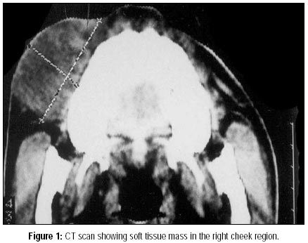
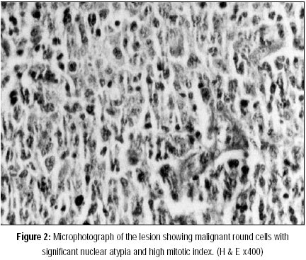
Journal of Postgraduate Medicine, Vol. 46, No. 3, July-September, 2000, pp. 211-212 Images in Medicine Primary Extra-nodal Non-Hodgkin's Lymphoma of the Cheek Maheshwari GK, Baboo HA, Gopal U*, Wadhwa MK** Departments of Radiation Oncology, Radio-Diagnosis* and Pathology** Code Number: jp00073 A 16-year-old non-smoker, non-tobacco addict boy presented with history of a slowly growing swelling over his right cheek region of 8-month duration with difficulty in opening the mouth for past 3 months. Local examination revealed a firm to hard, nodular subcutaneous mass on the right side of face reaching up to right lower jaw. The overlying skin was tense and shiny. Examination of oral cavity revealed mild trismus and 6x6x2 cm mass causing bulging over right buccal mucosa and gingivo-buccal sulcus region. The mass was well circumscribed and appeared to be free from the mucosa and bone. There was no cervical lymph node enlargement. Rest of the physical examination was normal. CT scan showed a mixed density mass in the right cheek region extending from the angle of mouth to retromolar region. There was loss of fat planes. The muscles of the cheek were involved but underlying jaw bones were free (Fig.- 1). The biopsy of the lesion on histopathological examination showed presence of submucosal tumour. The tumour cells were large in size, round in shape with hyperchromatic nuclei and high nuclear cytoplasmic ratio. Many tumour cells showed mitotic figures with few atypical mitoses. Intra-nuclear clefts, bi-nucleation and prominent nucleoli were also evident. Based on microscopic features (Fig. 2) possibility of poorly differentiated carcinoma or malignant lymphoma was suggested. Immuno-histochemical evaluation was performed that revealed positive immuno-reactivity for LCA (leukocyte common antigen) & PAN-B (CD-20). PAN-T, Cytokeratin (CK), AE1, Vimentin, S-100 and HMB-45 were negative thus, the final diagnosis of diffuse B-type large cell non-Hodgkin's lymphoma was established. The patient was evaluated for other sites of involvement and bone marrow aspiration, cerebrospinal fluid (CSF) examination & ultrasonography of the abdomen and pelvis were performed. There were no other sites in the body involved by the disease. The patient did not had B-symptoms. Thus, his disease was labelled as "I-E". The patient was initially subjected to systemic chemotherapy. He received 6 cycles of CVP (Cyclophosphamide, Vincristine, Prednisone) regimen. This was followed by local radiotherapy to the diseased area on 6 Mv linear accelerator using wedged anterior and lateral beams. He received a total dose of 50 GY/25 fractions/5weeks. Following the completion of treatment there was complete regression of the disease. He is currently on follow-up and remains disease free, 19-months after diagnosis and 13-months following completion of treatment.
Discussion Non-Hodgkin's lymphomas are a group of highly diverse malignancies and have great tendency to affect organs & tissues that do not ordinarily contain lymphoid cells. 20 to 30% of non-Hodgkin's lymphoma arise from extra-nodal sites. The head and neck is the second most common region for extra-nodal lymphoma after gastro-intestinal tract. Among various head & neck sites, Waldeyer's ring, which is area encompassed by the nasopharynx, the tonsil and the base of the tongue is most often involved by malignant lymphoma. The nose and para-nasal sinuses, orbit(s), salivary glands are other sites in head and neck affected in decreasing order of frequency. Involvement of the oral cavity is very uncommon. Among various intra-oral sites, the gingiva and palate are most often involved. Involvement of cheek or buccal mucosa, oral tongue, floor of mouth and lip(s) has been reported quite infrequently. Till date, only 26 cases of the non- Hodgkin's lymphoma of the cheek have been mentioned in the English literature.1-9 In Western countries, the incidence of carcinoma and non-Hodgkin's lymphoma of the oral region is roughly equal whereas in India, incidence of oral cancer involving the buccal mucosa or cheek is very high, but no case of non-Hodgkin's lymphoma of the cheek has been reported so far. Little is known about the aetiological factors for primary lymphoma of the oral region although, few cases of oral lymphomas have been reported in association with Acquired Immune Deficiency Syndrome, and it may even be the first indication of the disease in some individuals. Though it has been reported in all age groups, it generally affects the elderly, especially over the 6th decade of life.4 There are no characteristics clinical features of lymphoma of the oral region. The most common presenting symptoms are local swelling, pain or discomfort and ulcer. The oral non-Hodgkin's lymphoma may mimic more common benign oral and dental pathologic conditions.10 Thus, it may be mis-diagnosed. Awareness of this clinical entity in the oral region is important because lymphomas and more common malignant lesions such as carcinomas can not be differentiated clinically. Our patient had diffuse B-cell variety, large cell type non-Hodgkin's lymphoma. Most of the head and neck non-Hodgkin's lymphomas including oral lesions are of B-cell origin and diffuse large cell type being the most common.6 The paucity of cases makes the understanding of the biological behavior and therapeutic options of lymphoma of the oral region difficult. Like lymphomas at other head and neck sites, oral lesions also quite sensitive to both radiotherapy and chemotherapy. Our patient was treated with combined modality using chemotherapy and local radiotherapy because of high grade of tumour and large size of the lesion. The lesion responded well to treatment. He remains without evidence of disease 13-months past treatment. The overall prognosis of non-Hodgkin's lymphoma is related to the stage of tumour and the aggressiveness of the malignant cell type. In conclusion, though non-Hodgkin's lymphoma involving oral region is uncommon, it should always be considered in the differential diagnosis of various benign and malignant lesions in this region, because the treatment and prognosis for these conditions are quite different. A proper clinical examination and histopathologic as well as immuno-histochemical evaluation of biopsy specimen may aid in the diagnosis, and thus help in proper management.
References
This article is also available in full-text from http://www.jpgmonline.com/ Copyright 2000 - Journal of Postgradate Medicine The following images related to this document are available:Photo images[jp00073f2.jpg] [jp00073f1.jpg] |
| |||||||||
