 |
| About Bioline | All Journals | Testimonials | Membership | News |
|
||||||
|
||||||
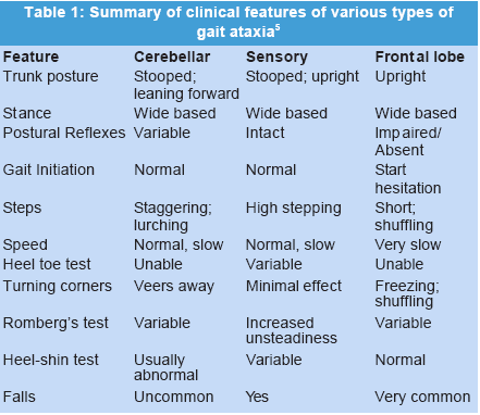
Journal of Postgraduate Medicine, Vol. 49, No. 2, April-June, 2003, pp. 169-172 Clinical Signs Romberg's Test Khasnis A, Gokula RM* Department of Internal Medicine, Michigan State University
and
*Department of Family Practice, Sparrow Health System, Lansing,
MI 48824, USA.
Code Number: jp03044 Summary Romberg's test is a simple bedside sensitive clinical test that pinpoints to sensory ataxia as the cause in a patient presenting with postural imbalance. As described above, the test is invaluable if carried out meticulously and interpreted cautiously. It must be carried out in all patients presenting with dizziness, imbalance, and falls. Sensory ataxia has a number of treatable etiologies. Needless to say that focused laboratory testing should be carried out in all cases to nail the cause and offer appropriate treatment. Romberg's test is a commonly performed test during the neurological examination to evaluate the integrity of dorsal columns of the spinal cord. Moritz Heinrich von Romberg first described it in the early 19th century. It evolved from a symptom to a valuable clinical sign. It was classically described in patients with tabes dorsalis (neurosyphilis), but can be elicited in many conditions affecting proprioception. This simple test offers an important clue to the presence of pathology in the proprioceptive pathway and should be meticulously carried out during the neurological evaluation. Early detection of reversible causes is desirable as they may be remediable and their treatment can prevent permanent dysfunction and disability. History In 1836, Marshall Hall first described a patient with tabes dorsalis who presented with complaints of increased unsteadiness in the dark. However, he did not develop this into a specific symptom or a sign.1 Ernst Horn, Romberg's teacher had five different students write their doctoral dissertations on this subject before the test was devised.2 In 1840, Moritz von Romberg developed the "symptom-presentation" into a test. Around the same time, Bernardus Brach noted similar symptoms in his patients. He also carefully noted that this imbalance was not due to muscular weakness. Unfortunately, his contributions are not as well recognized as those of Romberg and Hall are.1 In 1858, Duchenne de Boulogne reported cases of "progressive locomotor ataxia". He also observed that, in due course of their disease, patients with tabes dorsalis tend to lose their vision and this is accompanied by worsening ataxia.1 Tabetic ataxia was first described in detail by Duchenne in 1858. In 1921, Guillain described a reversible form of acute tabetic ataxia that develops abruptly and improves completely on early suitable treatment. Fournier described the early diagnosis of ataxia in detail and developed Fournier exercises (based on sudden and unforeseen movements).3 The importance of the test increased as the basis of abnormal findings was understood. Gradually it came to be used for excluding dorsal column abnormalities in patients presenting with unsteadiness of gait. Physiology of Proprioception4 Proprioception is a deep sensation that arises from the muscles, ligaments, tendons and joints. The peripheral organs (receptors) for proprioception are neuromuscular and neurotendinous spindles, Pacinian corpuscles and possibly Golgi tendon organs. These respond primarily to pressure, tension, stretching and related stimuli. Impulses from these receptors are carried by the large myelinated Ad fibers. The cell bodies of these neurons are located in the dorsal root ganglion (first order neuron). The nerve fibers carrying these impulses travel along the medial division of the dorsal root and ascend in the fasciculus gracilis and cuneatus till the nucleus gracilis and nucleus cuneatus (in the medulla) respectively where they synapse. The fascicul gracilis (tract of Goll) carries proprioception from the lower limb and lower trunk and lies medially in the dorsal columns, whereas the fasciculus cuneatus (tract of Burdach) carries it from the upper trunk and upper limb and lies laterally. Following decussation of the arcuate fibers in the medulla, the medial lemniscus carries this sensation to the contralateral ventral posterolateral nucleus of the thalamus. During their course, the fibers from the nucleus gracilis and cuneatus are anatomically related to each other as: ventral-dorsal (medulla), then lateral-medial (pons), dorsolateral-ventromedial (midbrain) and finally lateral-medial (thalamus). The impulses are then carried to the parietal cortex by the thalamoparietal fibers. These fibers terminate in the parietal cortex posterior to the fibers that convey touch. Joint position sense or sense of posture (also referred to as statognosis) refers to the awareness of the position of the body or its parts in space. Kinetic sense or sensation of active or passive movement (also referred to as arthresthesia) consists of awareness of motion of the various body parts. Patholophysiologic basis of Romberg test Central postural control (equilibrium) is dependent on input from three peripheral modalities: vision, vestibular apparatus and proprioception (joint sense and sense of position). Disturbance in any one of these modalities can be compensated for (completely or incompletely) by input from the other two systems. The dorsal columns in the spinal cord (fasciculus gracilis and cuneatus) are responsible for conducting proprioceptive, fine touch, pressure and vibratory impulses from the lower and upper limbs to the higher centers (brainstem, thalamus, and parietal cortex) for maintaining coordination. They can be affected by a variety of conditions ranging from subacute combined degeneration to tabes dorsalis. Impaired proprioception can be overcome by visual and vestibular feedback. However, reduced visual input in the dark surroundings or due to failing vision can seriously predispose such a patient to severe incoordination (ataxia). Asking the patient to close his eyes during the Romberg's test helps uncover any disordered proprioception that may have been masked by vision. Proprioceptive disturbances can arise at any level along the pathway described above. Various explanations have been proposed to explain why lack of visual input aggravates the imbalance - Change in limb muscle tone with active movement of the eyeballs, distraction of the patient's attention and suppression of visual spatial control when gaze is focused on a defined spot.3 The Romberg's test checks functional integrity of the entire proprioceptive pathway (tests for sensory ataxia). Although generally thought to be due to a disturbance of deep sensation, a positive Romberg's test has been described in patients with intact deep sensation. Jacoud and Marie3 have reported that in patients with tabes, the unsteadiness is less severe when prevented from seeing his own feet than when he closes his eyes. André-Thomas described that patients with tabes do not always need to look at their feet during walking; they often look straight ahead or focus on objects in front of them at a certain height. He also reported correction of imbalance even with eyes closed by training patients to use memory of a visual image of their body. He suggested that during clinical examination, the patient's attention is repeatedly aroused, which may help to sustain the input from the deep sensations; relaxing his attention may compromise the input. Interestingly, Benedikt has reported that the occurrence of blindness in tabes seems to prevent the development of ataxia; this is proposed to be due to an arrested pathological process (tabes fixé).3 These studies illustrate the problems with the Romberg's test and the interrelationship of vision, balance and voluntary movement. Clinical clues to a patient with ataxia: History As for other neurological disorders, a detailed history of a patient with ataxia is imperative and invaluable. Sensory ataxia should be differentiated from cerebellar, frontal lobe and vestibular ataxia. (Table 1) Only pertinent points in the history relevant to sensory ataxia are highlighted here. The onset, duration and progress of the ataxia should be noted. Sensory ataxia is usually insidious in onset. Sensory ataxia is usually more severe and worse if not exclusively present at night. A history of falling into the sink or imbalance when splashing water on the face (wash-basin sign), passing a towel over the face or pulling a shirt over the head should also be sought. The unsteadiness may show different features - staggering, instability, displacement of the trunk, flexion of the lower limbs and even a complete collapse. André-Thomas and De Ajuriaguerra have suggested that there is some relation between these features and the distribution of lesions in each case.3 History can also provide clues to the possible etiology: Diet (Pure vegetarian (vegan), devoid of any animal product including milk) and alcohol consumption (both predispose to vitamin B12 deficiency), ingestion of medications (vitamin B12 antagonists), diabetes (large fibre peripheral neuropathy), history of similar illness in other family members (hereditary causes of sensory ataxia), similar past episodes or past episodes of self-resolving blurring of vision (for multiple sclerosis), symptoms of cord compression (posterior lesion causing predominant dorsal column compromise), and history of unprotected sexual intercourse (for neurosyphilis). One should also remember to ask for symptoms of malignancy, as paraneoplastic neurological affection may be a cause for sensory ataxia. Accompanying changes in gait (stamping) and posture (stooping) should also be enquired about. Localization of the pathology may be aided by symptoms such as paresthesiae or "glove-stocking" anesthesia (peripheral nerves), involvement of bladder and bowel and affection of other sensory modalities (spinal cord), harlequin patterns of sensory disturbance (brainstem), accompanying persistent unilateral pain (thalamus) and accompanying higher cortical disturbances (parietal cortex). Complaints of loss of other modalities of sensation like fine touch, vibration and pressure also strongly favour large fibre neuropathy. Clinical clues to a patient with sensory ataxia: Physical examination During general examination, the patient must be examined for pallor (anemia), knuckle pigmentation (B12 deficiency), palpable thickened nerves (hereditary peripheral neuropathies), posture, hypo- and hyperpigmented patches on the skin, local examination of the spine (for scoliosis), presence of pes cavus (Friedrich's ataxia), and external telltale signs of internal occult malignancy. During neurological examination, higher functions must be tested (for neurosyphilis and dementia), the optic fundus must be examined (for pallor and degenerative changes), cranial nerves examined (especially CN II for visual acuity and Argyll-Robertson pupil, CN III, IV, and VI for ocular movements and CN VIII for vestibular function and deafness) and the motor system must be evaluated to rule out muscular weakness in the lower limbs as a cause of the ataxia. The sensory system must be tested meticulously for all sensations and special attention paid to fine touch, vibration and joint sense and position (large fibre carried sensory modalities). The technique and interpretation of the Romberg's test are detailed below. Sensory ataxia in the upper extremities is manifested as constant wandering of the outstretched upper limbs when the patient closes his eyes (pseudo-athetoid movements). The gait is an indispensable part of the examination in these patients. Patients with sensory ataxia have a "stomping" gait because they use the sensation of impact of the foot hitting the ground to guide their walk rather than touch or proprioception. The soles of the shoes of these patients are said to be uniformly worn-out as the entire plantar aspect of the foot strikes the ground at one time.6 Other clinical clues to differentiate the various types of ataxia are given in Table 1. Detailed examination for clinical evidence of neurosyphilis should be carried out. Finally, cerebellar signs should be looked for. Technique and interpretation of Romberg's test The patient should be examined to rule out other causes of the ataxia. The technique of the test should be explained to the patient. The patient is then made to stand with his feet close together, arms by the side and eyes open. Any significant swaying or tendency to fall is noted. The patient is then asked to close his eyes. Other maneuvers that may be used are looking away from the ground and asking the patient to follow the examiner's finger with his eyes as it moves rapidly from left to right or up and down.3 Postural swaying is again noted and compared with that observed with open eyes. The degree of swaying as well as its position should be noted (swaying from the ankles, hips or entire body). It is important to reassure the patient that he will be supported in case of severe imbalance. The physician should be facing the patient; his arms should be extended on either side of the patient to support him (without touching the patient). Romberg's test is considered positive if there is significant imbalance with the eyes closed or the imbalance significantly worsens on closing the eyes (if imbalance was present with the eyes open). Normal individuals also tend to sway to some extent on closing their eyes. Low normal performance consists of the ability to stand heel-to toe, with eyes closed, for six seconds. Young adults should be able to perform this test for thirty seconds but performance is reported to decline with age.7 Hysteria is an important cause of a false-positive Romberg's test. Patients with hysteria tend to sway from the hips rather than the ankles. However they do not fall and hurt themselves. In these patients, the imbalance can also be reduced by asking distracting questions or by doing the finger nose test simultaneously. Several procedures such as standing on one foot and one foot in front of the other have been suggested to increase the sensitivity of Romberg's test, but the test with the feet together is usually sufficient.3 Romberg's test is a simple test that can uncover sensory ataxia but must be interpreted with caution. Positive Romberg's test has also been reported in labyrinthine conditions, however Barré3 has pointed out that in this condition it is different from tabes. In tabes, the swaying begins as soon as the eyes are closed, occurs in all directions, and is rapid. In labyrinthine disorders, the imbalance appears after an interval, consists of a slow lateral inclination of the body, is of small amplitude, always in the same direction and may vary with the position of the head. Ataxia that is present with eyes open is suggestive of cerebellar aetiology. The other specific signs of cerebellar dysfunction should then be elicited. Lanska8 have reported use of quantitative, computer calculated Romberg's test and concluded that it is a reasonable way of measuring postural stability. They also suggest that Romberg's test has contributed significantly to the development of mechanical modalities like computerized dynamic platform posturography for measuring postural stability. Causes of positive Romberg's test Positive Romberg's test is a hallmark of sensory ataxia. However, a positive Romberg's test can result from: Inherited
Metabolic and toxic
Immunological and others
False positive Romberg's test
[Important and common causes have been mentioned in italics]. References
Copyright 2003 - Journal of Postgraduate Medicine. Online full-text also available at http://www.jpgmonline.com/ The following images related to this document are available:Photo images[jp03045t1.jpg] |
| |||||||||
