 |
| About Bioline | All Journals | Testimonials | Membership | News |
|
||||||
|
||||||
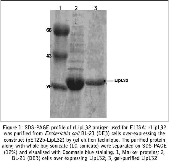
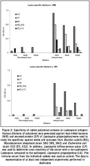
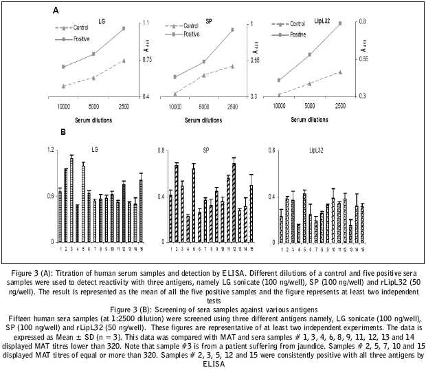
Journal of Postgraduate Medicine, Vol. 51, No. 3, July-September, 2005, pp. 164-168 Original Article Gel purified lipl32: A prospective antigen for detection of leptospirosis Tahiliani P, Kumar MMohan, Chandu D, Kumar A, Nagaraj C*, Nandi D Dept. of Biochemistry, Indian Institute of Science, Bangalore-560012 *Regional Office for Health and Family Welfare, Government of India, Kormangala, Bangalore-560034, India Code Number: jp05066 Related article: jp05067 Abstract Background: Leptospirosis, a zoonosis, is a re-emerging disease, affecting populations across the globe. However, the current methods of diagnosis are time- consuming, cumbersome, imprecise or expensive.Aim: To develop an assay for differential and early diagnosis of Leptospirosis. Methods and Material: IgG based ELISA for evaluation of three antigens, namely, a gel-purified recombinant protein (rLipL32), secreted proteins and whole organism sonicates of Leptospira spp. The antigens were evaluated using, rabbit polyclonal antiserum and human sera samples. Results: Studies with a rabbit polyclonal antiserum indicated the utility of these antigens in differentiating Leptospira from other common pathogenic organisms. Evaluation of these antigens with fifteen representative human serum samples indicated gel-purified rLipL32 to be a potentially useful antigen for detection of leptospirosis. The results obtained with IgG ELISA were correlated with the results of microscopic agglutination test (MAT). Conclusion: Gel-purified rLipL32 is a valuable antigen for early and accurate diagnosis of leptospirosis. Further evaluation of this assay in field conditions and larger sera samples will indicate its suitability in case of an epidemic. Keywords: Leptospira, LipL32, Human IgG ELISA, Gel elution Leptospirosis, an acute febrile illness, is a zoonotic disease caused by pathogenic spirochete of the genus Lept ospira.The disease, once considered to be an endemic or occupational/recreational hazard to people exposed to contaminated water, is now being recognised as a common cause of febrile illness in tropical environments worldwide. Often epidemics associated with high case fatality break out annually during periods of heavy rainfall in urban areas that lack basic sanitation facilities.[1],[2] Although, leptospirosis can be cured easily with antibiotic therapy, the confusion between the clinical presentation associated with leptospirosis and other febrile illnesses complicates the diagnosis. Symptoms of leptospirosis include high fever, severe headache, chills, hemorrhage, muscle aches, vomiting and may include jaundice, red eyes, abdominal pain, diarrhea or a rash, which are characteristic of a typical protean clinical presentation. If the disease is not treated in time, patients may develop renal damage, meningitis, liver failure, and respiratory distress; in rare cases death can occur[3],[4]. Thus, early and accurate diagnosis is a prerequisite for proper treatment of leptospirosis. The inherent limitations and intricacies of the presently available diagnostic methods, namely, culture, microscopic agglutination test (MAT) PCR and IgM ELISA further complicate the timely diagnosis of the disease. A lot of effort is being put to develop quick, simple and reliable methods of diagnosis for Leptospirosis. This study has been initiated to evaluate the usefulness of an indirect IgG ELISA for specific detection of anti-leptospiral antibodies in human patient serum samples. Methods and Materials Cloning, sequencing and over-expression of LipL32: LipL32 was PCR amplified from the thermolysate of Leptospira interrogans serovar grippotyphosa using gene-specific primers (forward 5′ctcccccgggatatgaaaaaacttcgattttggc3′ and reverse 5′ctcccccggggaatcctcaaagcttcttagtcgcgtcagaagc3′), cloned into pGEM ®-T cloning vector (Promega) and subcloned into pET22b expression vector (Novagen) using Nde I and EcoR I sites. Recombinant DNA techniques were performed essentially as described in Sambrook et al .[5] The primers were designed using the Leptospira interrogans sequence (available at www.ncbi.nlm.nih.gov) as template. The entire clone was sequenced using the automated DNA sequencing facilities at the Indian Institute of Science (IISc) or University of Delhi, South Campus. After sequence confirmation, the pET22b_LipL32 construct was induced and over-expressed in Escherichia coli BL21 (DE3) cells using lactose induction (1% lactose every 4 hours, induction carried out for a total duration of 8 hours). The medium was buffered with 10X phosphate buffered saline (PBS) prior to lactose induction to a final concentration of 1X to minimise pH alterations on lactose addition. Antigen preparation : Leptospira interrogans serovar grippotyphosa was obtained from Dr. Venkatesh, Institute of Animal Health and Veterinary Biologicals, Bangalore and was cultured in Ellinghausen-McCullogh Johnson-Harris (EMJH) medium at 30°C. Young cultures of the bacterium (approximately 10 days after inoculation), were centrifuged at 4,500 x g for 10 min to pellet the organisms. The pellet was washed twice with PBS to remove medium components, re-suspended in a small volume of PBS and incubated at room temperature for 12 hours. After incubation, the supernatant was collected by centrifuging cells at 10,000 x g for 10 minutes and used as secreted protein (SP). The protein content in the supernatant was measured using the Bradford assay.[6] The pelleted Leptospira were heat-killed (denoted as WB) by incubation at 80°C for 30 minutes thrice, with intermittent cooling at room temperature for two hours each time. The heat killed bacteria (WB) and secreted proteins (SP) from Leptospira interrogans serovar grippotyphosa were used to generate polyclonal antiserum using standard protocol and this serum was used to optimise the conditions for ELISA.[7] Organisms were sonicated at 20 kHz for 5 min., three to four times for use as an antigen (LG sonicate) in ELISA. The third antigen used in this study, rLipL32 was purified by gel elution. In short, cells over-expressing the pET22b_LipL32 construct were harvested by centrifuging the culture at 5500 X g for 10 min. The cells were sonicated and centrifuged at 7000 X g for 20 minutes to remove the cell debris. The supernatant thus obtained was centrifuged again at 10,000 X g for 2 hours to remove residual cell debris. The supernatant was then loaded on preparative gels (12% SDS-PAGE); after completion of the run, a small strip of the gel was cut and stained with Coomassie Blue to visualise protein bands. The LipL32 band was cut from the unstained portion of the gel, washed with PBS twice to remove excess SDS adhering to the gel, and was crushed into small pieces. The protein was eluted by incubating gel pieces in PBS at 4°C overnight on an end-to-end rotor. The eluate was concentrated using Amicon membrane concentrators with 10 kDa cut off (Millipore Corporation, USA), checked for protein concentration and confirmed by SDS-PAGE. This was further used as gel eluted LipL32 antigen in the IgG-ELISA. The quality of antigen was reproducible as assessed with the rabbit antiserum as well as with human samples. In fact, the ELISA data represented in this study were performed minimum three times and with at least two independent antigen preparations. Indirect IgG-ELISA : Polyvinyl chloride microtiter plates (Corning Incorporation, USA) were coated with appropriate antigens in varying concentrations (0.05 - 0.1 µg/well) in 50 mM PBS. The plates were incubated at 37°C for two hours. Unbound antigens were washed off with PBST (50 mM PBS containing 0.2% Tween-20). Further, the wells were treated with 1% fraction V-bovine serum albumin (BSA) (Sigma-Aldrich Co., USA) in 50mM PBS at 37°C for 2 hours. The excess BSA was washed off and 100 µl of various serum dilutions, PBS and control serum were added to appropriate wells. The antigen-antibody reaction was allowed to take place at 37°C for 1 hour. After washing the wells thoroughly with PBST, secondary antibody (peroxidase conjugated AffiniPure Goat Anti-Human IgG (H+L) from Jackson Immunoresearch laboratories INC.) at a dilution of 1:5000 (100 µl/well) was added for 1 hour. The plates were washed thrice with PBST followed by washing thrice with PBS. The reaction was developed by incubating TMB/H2O2 substrate (20X solution from Bangalore Genei, India) prepared in distilled water. The reaction was stopped with 2N H2SO4 and the plate was read at 450 nm using an ELISA plate reader (Molecular Devices, USA). Human patient samples : Fifteen different sera samples representing varied patients (suspected to be suffering from leptospirosis) were picked up randomly from a pool of samples, which came for routine investigations. The control serum sample was from a healthy person. The serum samples were then evaluated against all the three antigens using the IgG-ELISA. Results Lactose induction of the cells transformed with the pET22b_LipL32 construct resulted in over-expression of LipL32, as detected by SDS-PAGE in lane 2 [Figure - 1]. Gel elution of rLipL32, from extracts of cells over-expressing rLipL32 after separation on SDS-PAGE, resulted in a single protein ( > 95%, based on Coomasie Blue staining) which showed the same mobility on SDS-PAGE in lane 3 [Figure - 1]. The polyclonal antiserum generated against heat-killed Leptospira (top) and the secreted protein (bottom) was specific in detecting Leptospira and did not cross react with other microbes tested, for example, Bacillus subtilis , Escherichia coli , Mycobacterium smegmatis [Figure - 2]. The data in [Figure - 2] demonstrate that the antisera generated against the Leptospiral antigens was specific and did not crossreact with other microbial antigens. Further, these antisera were used to optimise the conditions of ELISA. ELISA results generated by the screening of the three antigens with human serum samples indicated that gel-purified rLipL32 detected Leptospirosis with high specificity (80% at a cut off of Mean+2SD), which is more than that of secreted protein (66%) and LG sonicate (73%). Similarly, rlipL32 was more sensitive (80%) when compared to both SP as well as LG sonicate (60%) in detecting IgG antibodies against Leptospira in these patients [Figure - 3]. The selection of the cut off values for ELISA were determined using the control human serum sample (#1) [Figure - 3]B.The borderline (OD) was 1.5 times the OD of the control serum under identical test conditions. As some fluctuations in the background OD values were observed from experiment to experiment, only the sera that were consistently positive in independent experiments (n = 3), were considered to be truly positive by ELISA. These data were considered together with MAT data to calculate specificity and sensitivity. Leptospira interrogans serovar pomana was used as antigen in Micro Agglutination test [MAT]. Sera that tested positive by MAT had titres of 320 going upto more than 5120. The MAT positive serum that was negative by ELISA may be having very low titre of IgG antibodies. Patients who displayed titres of 320 and above were diagnosed as suffering from Leptospirosis based on: (i) Clinical picture (ii) Dark ground microscopy of MAT positive sera and (iii) clinical response with penicillin. Discussion The key factor for the proper treatment of leptospirosis is early and accurate diagnosis. Though there are many methods available for its diagnosis by detection of the pathogen, they are tedious, time-consuming and are not very specific, as most of them detect mainly agglutinating IgM antibodies (either by the formation of agglutinations or in ELISA format). The main drawback with detection assay based on these antibodies is lack of specificity, as IgM antibodies are known to be cross-reactive and form agglutination complexes with antigens from across the bacterial species. MAT requires a battery of representative live organisms for proper diagnosis; maintaining these organisms is not easy and requires skilled personnel and specialised facilities. Therefore, an IgG antibody based assay system was adopted in ELISA format for specificity and ease of detection. When rabbit polyclonal antiserum was used to optimise the conditions for IgG-ELISA, it showed linear titration with varying dilutions and significant positive values were observed even at the dilution of 1:10000, indicating the high sensitivity of the assay system. Further, when the antiserum was used to study cross-reactivity against secreted proteins from other common bacteria ( Escherichia coli, Mycobacterium smegmatis and Bacillus subtilis ), it was found to be specific for leptospiral proteins. Interestingly, at higher dilution (1:5000, 1:10000) the polyclonal serum was also able to detect only pathogenic Leptospira as against non-pathogenic Leptospira biflexa serovar patoc. The polyclonal serum generated against secreted proteins from Leptospira grippotyphosa was more specific in differentiating the pathogenic organism from non-pathogenic members. These results led us to use a purified protein from the pathogenic Leptospira to increase the specificity of the detection system. In-silico analysis (BLAST search followed by multiple sequence alignment using ClustalW) using sequence of LG-LipL32 as template revealed that this lipoprotein was unique to pathogenic Leptospira. In fact, no significant orthologs were detected in complete genome sequences of other organisms (data not shown). The alignment also indicated that the protein is polymorphic. LipL32 is highly conserved in pathogenic Leptospira across the globe, as evidenced by the LipL32 sequences submitted from various parts of the world, further highlighting the effectiveness of the protein for use as an antigen in ELISA, specific for detection of leptospirosis, as also suggested by earlier works.[8] Thus, we resorted to the use of purified rLipL32 for developing the specific IgG ELISA and to validate it in principle for diagnosis of Leptospirosis in human serum samples. Evaluation of gel-purified rLipL32 based IgG-ELISA with human serum samples also gave the same results as rabbit antiserum. The presence of minor amounts of SDS in the antigen preparation did not interfere with the antigen-antibody reaction (data not shown) in keeping with the earlier reports suggesting the use of SDS-purified ligands in ELISA.[9] The results of human IgG-ELISA were in accordance with the results of MAT, using only Leptospira interrogans serovar pomana as antigen. Among the three antigens, rLipL32 was found to be the most sensitive and the most specific in detecting leptospirosis. The sensitivity and specificity (both 80%) of rLipL32 based IgG ELISA was better than those of sonicate and the secreted proteins. Further, the use of a gel-purified recombinant protein overcomes the usual drawbacks of batch to batch growth variation in the preparation of sonicates of whole organisms and is also more convenient and cheap compared to the use of purified proteins where purification of the protein is a cumbersome process in itself and also requires a lot of infrastructure thereby increasing the cost of assay. Although, results of this study suggest the potential of this antigen for differential and accurate diagnosis of leptospirosis in principle, further research is necessary to evaluate the effectiveness of this antigen with a larger number of samples during outbreaks of Leptospirosis. The common criticism of using an IgG based ELISA for early diagnosis is the relatively late appearance of these antibodies during the course of immune response against any antigen. However, it has been shown that the kinetics of IgG response against leptospiral recombinant proteins is comparable to the IgM response against whole antigen preparations[8],[10] Further, it was shown that the recombinant protein was able to induce a robust IgG response against an undetectable IgM response, which was presumably due to faster IgM-IgG seroconversion.[8],[10] Moreover, LipL32 is known to be the most abundant outer membrane protein, which is expressed during all mammalian infections and reacts with patient serum from all phases of infection further pointing to its usefulness as an antigen for specific detection of leptospirosis.[11],[12],[13] This study demonstrates the usefulness of a recombinant antigen directly purified from PAGE, resulting in a much easier purification strategy compared to an earlier report.[11] In addition to previous reports of LipL32 as a potential antigen,[10],[11] the results of this study confirm the use of LipL32 as a possible diagnostic antigen. This study makes a prima-facie case for the use of gel-purified rLipL32 based IgG-ELISA for screening human patients for the detection of Leptospirosis. Further evaluation of this test is required with a larger sample set especially when there is an epidemic. Acknowledgement We are grateful to Lt. Gen. D. Raghunath for encouraging this study and for his comments on this work. We thank Dr. Venkatesh, IVB&AH, Hebbal, Bangalore for providing leptospiral isolates. A Post Doctoral Fellowship to PT from DBT is gratefully acknowledged. The financial support by the Sir Dorabji Tata Centre for Research in Tropical Diseases, Bangalore is greatly appreciated.References
Copyright 2005 - Journal of Postgraduate Medicine The following images related to this document are available:Photo images[jp05066f1.jpg] [jp05066f3.jpg] [jp05066f2.jpg] |
| |||||||||
