 |
| About Bioline | All Journals | Testimonials | Membership | News |
|
||||||
|
||||||
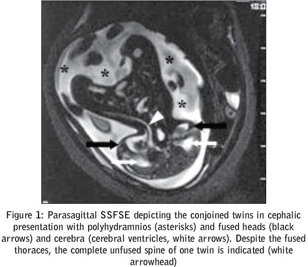
Journal of Postgraduate Medicine, Vol. 51, No. 3, July-September, 2005, pp. 228-229 Images In Radiology Ultrafast magnetic resonance imaging of cephalothoracopagus janiceps disymmetros Khanna ParitoshC, Pungavkar SA, Patkar DP MRI, Department, Nanavati Hospital, Mumbai Code Number: jp05082 Conjoined twins, rare variants of monozygotic twins, result from an incomplete division of the embryonic disk.[1] Conjoined twinning occurs once in every 50,000-100,000 births. Cephalothoracopagus, a very rare variant of conjoined twins occurs once in every three-million births.[2] Early prenatal diagnosis is desirable, given the poor prognosis.[1] A 28-year-old primigravida at 24-weeks gestation underwent a routine obstetric ultrasonography that revealed a foetus with an unusually large head and thorax, two separate heart pulsations and multiple limbs. While conjoined twinning was considered, the exact anomaly was difficult to elucidate. There was no history of consanguinity, previous twinning, diabetes, hypertension or ovulation-induction and any other drug use. The femur length was 5.2 cm. A MRI of the foetus [Figure - 1] was performed on a 1.5-Tesla unit. A six-element phased-array torso coil was used without maternal sedation or foetal curarization. Single Shot Fast Spin Echo (SSFSE) sequences were performed in multiple planes. The gravid uterus was scanned in three orthogonal planes. A monochorionic, monoamniotic gestation with Janiceps conjoined male twins was noted, having a single fused head with two complete faces and four eyes, duplicated, frontally fused cerebra with unfused spines, a fused thorax with two hearts, and eight limbs. The couple was counseled and they opted for pregnancy termination by induction. An autopsy confirmed the diagnosis of cephalothoracopagus janiceps. Each foetus had normal internal thoracic, abdominal and pelvic organs. Discussion We present a case in which magnetic resonance imaging (MRI) was employed for the antenatal diagnosis of a 24-week cephalothoracopagus foetus. Janiceps (jan·i·ceps) [Latin; Janus two-faced god + caput, head] are conjoined twins with one head and two faces looking in opposite directions [Figure - 2]. Janiceps are non-viable embryos with cephalic elements that are frontally fused. The developing facial regions form laterally into a compound structure halfway between the embryos, resulting in a pair of ′lateral′ faces, which are thus composed of two ′hemifaces′, each belonging to one embryo. The somatic axes are in the same plane and in opposite directions. These two different symmetry axes make this anomaly a valuable tool to study midline structure and optic commissure formation in humans. As described by Viggiano D et al and exemplified by our case, the optic nerves on MRI appeared to converge toward a distorted sphenoid bone. Two optic commissures were noted at autopsy, each in connection with a face, thus respecting the face midline and not the brain midline.[3] Janiceps ′monosymmetros′ or ′asymmetrus′ are terms used for variants that have a single hypoplastic face that is composed partially from the face of one twin and partially from that of the other and imply ′asymmetry′ or ′imbalance′.[1],[2],[4] Often, two dissimilar faces are seen with one showing incomplete facial features such as a single naris, cyclopia, synotia, a proboscis or two small eyes in a single palpebral fissure.[2] Janiceps ′disymmetros′, a variant with two identical, symmetrical faces is unlike Janiceps Asymmetros , in that the two notochordal axes are oriented in a perfectly ventroventral fashion. As in our case, the two faces were on opposite sides of the head and were identical and normal-looking.[2] The earliest antenatal diagnosis of cephalothoracopagus twins reported in the literature was made by vaginal ultrasound at eight-weeks gestation. A false-positive result in the first trimester may result from the close proximity of the foetuses.[2] Therefore, a definitive diagnosis with conventional ultrasound is generally made only in the early third trimester, when a detailed assessment of the degree of fusion can be made.[2],[4] 3D ultrasound employed at ten-weeks gestation can generate accurate, surface-rendered images of the twins assessing the anatomical relationship of the craniofacial structures, foetal skeleton, spine and thorax.[1],[2] 3D power Doppler can be used for vascular anatomy mapping at the conjoined site.[2] Post-mortem radiography and 2D or 3D multi-detector row computed tomography (CT) are supportive.[2],[5],[6] Our case was conclusively diagnosed with MRI. A classical cephalothoracopagus janiceps disymmetros foetus with frontally fused cerebra, separate spines, identical faces and four limbs each, was well demonstrated. Antenatal MRI in the past was not a suitable alternative to ultrasonography, due to the long scan time. Technological advances and faster sequences allow acquisitions to be completed in the interval between foetal movements. Subtle anomalies are detectable with MRI due to its high spatial and temporal resolution, multiplanar capabilities and superior soft-tissue characterisation. MRI allows a wider field-of-view, permitting a more complete foetal evaluation. From an obstetric standpoint, prenatal diagnosis of shared organs dictates possible surgical separation or pregnancy termination strategies. Generally, before 24-weeks, termination of pregnancy by the vaginal route with destructive procedures is employed; after this, termination by hysterotomy is considered prudent due to the potential for dystocia.[7] Acknowledgement The authors thank Punita Khanna for manuscript text and image editing.References
Copyright 2005 - Journal of Postgraduate Medicine The following images related to this document are available:Photo images[jp05082f2.jpg] [jp05082f1.jpg] |
| |||||||||
