 |
| About Bioline | All Journals | Testimonials | Membership | News |
|
||||||
|
||||||
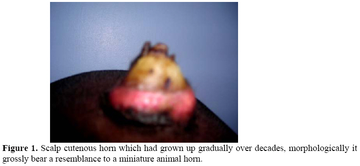
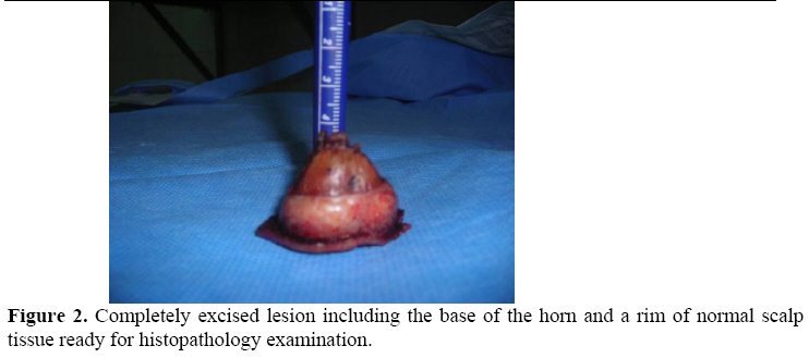
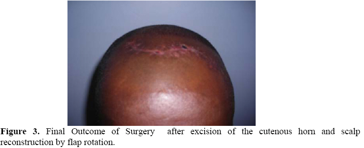
East and Central African Journal of Surgery, Vol. 13, No. 2, September, 2008, pp. 83-86 Giant Scalp Cutaneous Horn In Black Malawian Adult: A Case Report S.J. Uledi, M.B. Kalengayi Department of Surgery Zomba Central Hospital, Zomba, Malawi. Code Number: js08037 Background: Cutenous horn is a common disease entity in European Caucasians and other populations in Asia, far-east, and North America. By far reported cases involving black Africans are exceedingly few. This presentation therefore is aimed to yet document another occurrence of a rare case from Malawi involving an adult black African male with scalp cutenous horn. Also to sensitise Clinicians in our environment to be aware of the fact that cutenous horn does exist in black African populations, and should be considered when evaluating patients with dermatological conditions. This is a case description, including clinical and histopathological features of a patient seen at Zomba Central hospital, south east Malawi in April 2008. Introduction The term cutenous horn or cornu cutaneum is a clinical diagnosis referring to a morphological description of a solid outgrowth or conical projection above the surface of the skin that bear a resemblance to a miniature animal horn1. This condition is relatively rare clinical entity to find in black Africans, and when encountered may pose a diagnostic challenge to a clinician. It is only very recently that probably the first typical case of a cutenous horn in black African was reported by the Kenyan surgeon2. Prior to this, there was only one published report from northern Sudan where large segment of the population is of an African –Arab origin3 . Despite the fact that these lesions have been less frequently reported in our environment their existence is well known world over. The earliest observations on cutenous horns in humans were described as far back as seventeenth century by the London surgeon Everard Home4.To date, several and different forms of cutenous horns have been described mainly involving European Caucasians and other populations in Asia, Far east and North America4-6. Cutenous horns most often occur in adults, usually elderly, fair skinned individuals with history of significant sun exposure. The most affected areas include, scalp, face pina, nose, eyelids, neck, shoulders, forearms and dorsal hands. However some lesions may occur even on sun protected areas like nostrils and penis 7, 8. These horns may originate from a variety of benign, premalignant, or malignant epidermal lesions. Therefore histopathology examination specifically of the base is of paramount importance to rule out associated malignancy4-6 . This presentation is aimed at reporting yet another seldom case of cutaneous horn from Malawi (South-Central Africa) in an adult male black African. In one way, this may once again remind us on the existence of this rather rare disease entity in black Africans, as well as ensuring that no interesting and educative pathologies goes under reported. Case Report A 43 years old Malawian male with dark African complexion from Taombe village, Zomba district, south east Malawi. Presented to Zomba central hospital surgical outpatient department in April 2008, with long standing history of gradual onset of a painless but progressively growing swelling on the frontal aspect of the scalp for the past 28 years. He reported to have been relatively well until about 28 years ago when he first noted a painless small solid scalp eruption on the frontal aspect of the scalp. Initially, being a very small swelling, covered with hair, painless and growing at negligible rate the patient did not pay heed to it. However with time and in particular the last five years prior to his presentation, the swelling started growing much faster and became readily noticeable, a situation that somewhat caused social stigma and compelled him to start seeking medical attention. Before coming to Zomba hospital, patient reports to have visited several peripheral health facilities where by he was put on a wide range of antibiotics and many topically applied remedies with no improvement. He also reports to have consulted a number of traditional healers with no success. Associated with the scalp swelling patient gave no history suggestive of any neurological deficits or other significant concomitant symptoms. The patient, a peasant farmer by occupation mainly grews maize, cassava and sweet potatoes. He reported to have spent on average about eight hours a day, farming without sun protection gears. His wife and 4 children were all alive and well. He gave no history suggestive of skin cancer in the family. He neither took alcohol nor smoked tobacco in any form. On examination, the significant findings were on local examination, however on general examination, we saw a middle aged black African male in good nutritional status, well oriented, afebrile with no cervical or occipital lymphadenopathy. Locally, there was a cone-shaped mass arising from the frontal region of his scalp about 3cm from the fore head hair-line. The size of the swelling was 3.5cm (Base diameter) x 3cm (Width) x 2.5cm (Height) in, with well circumscribed margins. The base was encircled with an area of hypopigmentation but the rest of the scalp skin was normal. Its peak was surmounted with a conical hard keratotic protuberance that largely resembles a miniature animal horn. This horn was golden-yellow in colour (Fig 1). On palpation, the swelling had a firm consistency at the base and it was non tender, its peak which was surmounted with the horn, felt hard. The swelling was not fixed to the underlying structures and it was fairly mobile in all planes. Other systemic examinations were unremarkable. The lesion was excised with a rim of normal tissue and sent for histopathology examination (Figure 2). Primary closure was achieved by doing flap rotation and patient had uneventful recuperation (Figure 3). Histology specimen showed compacted keratin lying on a cratenformbase with papillomatosis and cutaneous horn formation. No evidence of malignancy was detected. Discussion Cutenous horns may arise from any part of the body and may vary considerably in size and shape. Like in our case, cutenous horns more often occur singly and may grow slowly over decades9. Although macroscopically cutenous horns may largely appear like miniature animal horn, histologically are quite different, human horn lacks centrally positioned bone10. In our case the patient had skull x-ray done as part of the base line investigations prior to excision but the lesion was radiolucent Cutenous horns are common in older and light-skinned individuals; this probably explains why they are less frequently encountered in our environment. The higher predilection in older and light –skinned individuals is grossly ascribed to the fact that many cutenous horns are caused by cumulative ultraviolet light damage over many years leading to actinic keratoses and non melanoma skin cancer6,11. Usually the peak occurrence of cutenous horn is in persons aged 60 years to 75years1,11,12. In the case that was reported from Kenya and this report from Malawi patients are far much younger, aged 28 years and 43 years respectively, though both had their horns growing slowly over years. Cutenous horns are predominantly benign lesions (60%)6,7. Up to 40% of cutenous horns may occur as part of premalignant or malignant skin lesions. No clinical features reliably distinguish between benign and malignant lesions. There fore histopathology confirmation is often necessary to rule out malignant changes. Features that have been reported to increase the chance of underlying malignancy includes older age, male sex, lesion geometry (either a large base or a large height to base ratio ) and presence on a sun exposed location1,12. In this particular case, histological findings revealed no underlying malignancy; this to some extent is reflected by the asymptomatic long standing history and slow growth of the lesion for about 28 years. We believe that in our case, the sun exposure factor could have played a major role as an important etiological factor in the pathogenesis of this lesion. Treatment modalities of these lesions are contingent upon the type of lesion at the base. Surgical excision that includes the base of the horn and appropriate margins with careful histological examination to exclude focus of malignancy remains the treatment of choice. Conclusion This is yet another seldom case of cutenous horn reported from Malawi (South-Central Africa) involving an adult male black African. The report reminds us once again on the existence of this rather rare disease entity in black Africans. The general objective of this report is to sensitise and make clinicians in our environment aware of the fact that cutenous horn does exist in black African populations and should be thought of when evaluating patients with dermatological conditions. References
© 2008 East and Central African Journal of Surgery The following images related to this document are available:Photo images[js08037f1.jpg] [js08037f3.jpg] [js08037f2.jpg] |
| |||||||||
